Search results (102 results)
-
 Lady in a dress
Feb 9 2023 by Shelby Helton
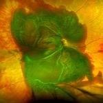
Fluorescein Angiography on a 67-year-old male with significant RPE changes secondary to a severe subretinal hemorrhage that required a vitrectomy with subretinal TPA in 2013.
Photographer: Shelby Helton
Imaging device: Heidelberg Spectralis
Condition/keywords: wet age-related macular degeneration (wet AMD)
-
 Massive SRH in Patient on Coumadin Being Treated for Exudative AMD
Sep 30 2019 by John S. King, MD
78-year-old white female using 1mg of warfarin for atrial fibrillation, who had a large PED, Type 1 lesion from AMD. Noticed acute darkening of vision one week after anti-VEGF injection. Has very large SRH, subRPE heme, and corrugated retinal appearance post RPE tear. Vision HM (from 20/100). 20/25 in the fellow eye that has dry AMD.
Photographer: Shelly Blair
Imaging device: Optos CA
Condition/keywords: subretinal hemorrhage, wet age-related macular degeneration (wet AMD)
-
 RPE-Transplantation
Jul 25 2024 by Gabriel Costa Andrade, PhD
Postoperative period of RPE-transplantation in a patient with neovascular AMD after RPE tear.
Photographer: Gabriel Andrade
Condition/keywords: neovascular age-related macular degeneration (AMD), pars plana vitrectomy (PPV), wet age-related macular degeneration (wet AMD)
-
 Chronical Submacular Hemorrhage in the Setting of Neovascular AMD
Mar 23 2015 by Rita Couceiro, MD, MS
An 80-year-old male, with a history of hypertension and high cholesterol, complained of acute and painless vision loss in his left eye (OS) in the previous 5 months. On observation best corrected visual acuity in OS was hand motion. A dense vitreous opacity in OS precluded fundus examination. Ocular ultrasound revealed vitreous hemorrhage and thickening of the macular area. The patient was submitted to pars plana vitrectomy, which disclosed a large submacular hemorrhage with chronical features and disciform scarring in the setting of neovascular AMD.
Imaging device: Intraoperative fundus photograph
Condition/keywords: neovascular age-related macular degeneration (AMD), submacular hemorrhage, wet age-related macular degeneration (wet AMD)
-
 RAP Lesions
Sep 29 2014 by Thomas A. Ciulla, MD, MBA, FASRS
Fluorescein angiogram of an 81-year-old man revealing several RAP lesions superior to fovea.
Photographer: Stuart Alfred CRA
Condition/keywords: choroidal neovascular membrane (CNVM), neovascular age-related macular degeneration (AMD), retinal angiomatous proliferation (RAP), wet age-related macular degeneration (wet AMD)
-
 Choroidal Hemorrhage, Subretinal Hemorrhage
Dec 18 2017 by Nichole Lewis
Choroidal hemorrhage, Subretinal Hemorrhage, wet macular degeneration,
Photographer: Nichole Lewis
Condition/keywords: choroidal hemorrhage, choroidal neovascularization (CNV), exudative age-related macular degeneration, subretinal hemorrhage, wet age-related macular degeneration (wet AMD)
-
 Exudative Macular Degeneration
Apr 26 2019 by Pamela Rowlett
Exudative macular degeneration
Photographer: Pamela Rowlett CRA, Universtiy of Colorado, Rocky Mountain Lions Eye Institute
Condition/keywords: wet age-related macular degeneration (wet AMD)
-
 Subretinal Hemorrhage Due to SRNVM, Fluorescein Angiogram Photograph
Dec 1 2016 by James B. Soque, CRA, OCT-C, COA, FOPS
89-year-old white male with NVAMD and new subretinal hemorrhage, fluorescein angiogram, early phase, of the right eye. Currently receiving anti VEGF treatment.
Photographer: James Soque, CRA, OCT-C, COA, Island Retina, Shirley, NY
Imaging device: Topcon TRC 50 DX, with MERGE software
Condition/keywords: hemorrhage, Hot spot, neovascular age-related macular degeneration (AMD), subretinal hemorrhage, subretinal blood, wet age-related macular degeneration (wet AMD)
-
 Active Neovascular AMD With Disciform Scar
Apr 30 2015 by Mitzy E Torres Soriano, MD
Active neovascular AMD with disciform scar.
Photographer: Mitzy E. Torres Soriano, MD; Centro medico Cagua-Estado Aragua. Venezuela
Imaging device: TOPCON
Condition/keywords: disciform scar, disciform with hemorrhage, neovascular age-related macular degeneration (AMD), wet age-related macular degeneration (wet AMD)
-
---thumb.JPG/image-square;max$300,300.ImageHandler) Advanced wet AMD
Advanced wet AMD
Dec 1 2013 by Mallika Goyal, MD
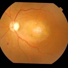
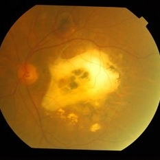
Right eye of a 77 year old male patient with a large RPED, sub neurosensory retinal fluid and submacular bleed following 10 months gap in anti-VEGF therapy.
Photographer: Mallika Goyal, MD, Apollo Hospitals, Hyderabad, India
Condition/keywords: wet age-related macular degeneration (wet AMD)
-
---thumb.JPG/image-square;max$300,300.ImageHandler) Advanced Wet AMD
Advanced Wet AMD
Jan 29 2014 by Mallika Goyal, MD
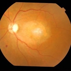
Right eye of a 77-year-old male patient with bilateral advanced wet AMD shows regressing RPE detachment following 3 years of anti-VEGF therapy.
Photographer: Mallika Goyal, MD, Apollo Health City, Hyderabad, India
Condition/keywords: wet age-related macular degeneration (wet AMD)
-
---thumb.JPG/image-square;max$300,300.ImageHandler) Advanced Wet AMD
Advanced Wet AMD
Jan 29 2014 by Mallika Goyal, MD
Left eye of a 77-year-old male patient with bilateral advanced wet AMD shows regressing RPE detachment and extensive scarring following 3 years of anti-VEGF therapy.
Photographer: Mallika Goyal, MD, Apollo Health City, Hyderabad, India
Condition/keywords: wet age-related macular degeneration (wet AMD)
-
 Advanced Wet AMD
Jan 29 2014 by Mallika Goyal, MD
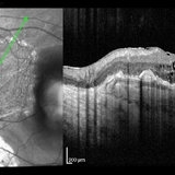
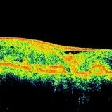
OCT of the right eye of a 77-year-old patient with bilateral advanced AMD shows massive RPE detachment and sub sensory retinal fluid at presentation in January 2011.
Photographer: Mallika Goyal, MD, Apollo Health City, Hyderabad, India
Condition/keywords: wet age-related macular degeneration (wet AMD)
-
 Advanced Wet AMD
Jan 29 2014 by Mallika Goyal, MD
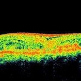
OCT of the right eye of a 77-year-old patient with bilateral advanced AMD shows regressing RPE detachment 1 year after initiating anti-VEGF therapy.
Photographer: Mallika Goyal, MD, Apollo Health City, Hyderabad, India
Condition/keywords: wet age-related macular degeneration (wet AMD)
-
 Advanced Wet AMD
Jan 29 2014 by Mallika Goyal, MD
OCT of the right eye of a 77-year-old patient with bilateral advanced AMD shows regressing RPE detachment 3 years after initiating anti-VEGF therapy.
Photographer: Mallika Goyal, MD, Apollo Health City, Hyderabad, India
Condition/keywords: wet age-related macular degeneration (wet AMD)
-
 Advanced Wet AMD
Jan 29 2014 by Mallika Goyal, MD
OCT of the right eye of a 77-year-old patient with bilateral advanced AMD shows regressing RPE detachment 2 years after initiating anti-VEGF therapy.
Photographer: Mallika Goyal, MD, Apollo Health City, Hyderabad, India
Condition/keywords: wet age-related macular degeneration (wet AMD)
-
 Advanced Wet AMD With Epimacular Membrane
Jul 15 2014 by Mallika Goyal, MD
Left eye of an 80-year-old lady with diminished vision for 1 year shows prominent epimacular membrane with underlying advanced wet AMD lesion.
Photographer: Mallika Goyal, MD, Apollo Health City, Jubilee Hills, Hyderabad-500033
Condition/keywords: wet age-related macular degeneration (wet AMD)
-
 Advanced Wet AMD with Epimacular Membrane
Jul 15 2014 by Mallika Goyal, MD
Left eye of an 80-year-old lady with diminished vision for 1 year shows prominent epimacular membrane with underlying advanced wet AMD lesion.
Photographer: Mallika Goyal, MD, Apollo Health City, Jubilee Hills, Hyderabad-500033
Condition/keywords: wet age-related macular degeneration (wet AMD)
-
 Advanced wet AMD with scar and persisting activity
Jul 15 2014 by Mallika Goyal, MD
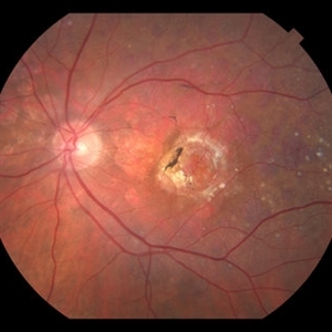
Left eye of a 76-year-old male with bilateral large macular scars from wet AMD. This eye has a new active lesion superior to the macular scar and fluid in the area of the scar suggestive of ongoing activity.
Photographer: Mallika Goyal, MD, Apollo Health City, Jubilee Hills, Hyderabad-500033
Condition/keywords: wet age-related macular degeneration (wet AMD)
-
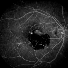
 Advanced Wet AMD With Scar and Persisting Activity
Jul 15 2014 by Mallika Goyal, MD
Left eye of a 76-year-old male with bilateral large macular scars from wet AMD. This eye has a new active lesion superior to the macular scar and fluid in the area of the scar suggestive of ongoing activity.
Photographer: Mallika Goyal, MD, Apollo Health City, Jubilee Hills, Hyderabad-500033
Condition/keywords: wet age-related macular degeneration (wet AMD)
 Loading…