Search results (27 results)
-
 Posterior Vitreous Detachment
Sep 1 2020 by J. Sebag, MD, FACS, FRCOphth, FARVO
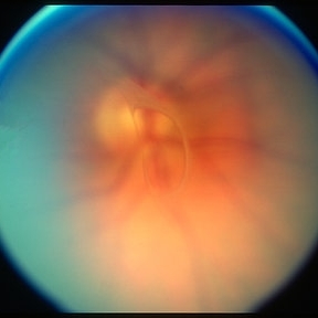
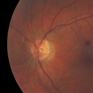
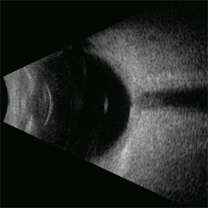
Left: Preset lens biomicroscopy of PVD in the left eye of a subject with a widely dilated pupil. The detached posterior vitreous cortex is seen (arrows) as is the optic disc and retinal vasculature (upper left). (courtesy of C. L. Trempe MD, Harvard Medical School, Boston, MA) [Sebag J: Vitreous – in Health & Disease Springer, New York, 2014; image © Springer Nature, reprinted with permission] Right: B-scan ultrasonography of PVD images the detached posterior vitreous cortex with a visible Weiss Ring.
Condition/keywords: posterior vitreous detachment
-
 Posterior vitreous detachment
Jan 11 2013 by Alex P. Hunyor, MD
Posterior vitreous detachment with prominent Weiss ring.
Condition/keywords: posterior vitreous detachment
-
 Weiss Ring
Jan 9 2019 by John S. King, MD
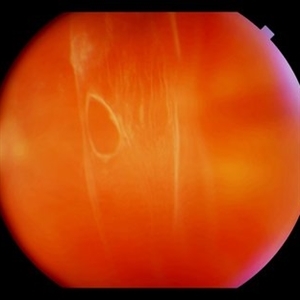
77-year-old white male with ERM and PVD OD; sheet of vitreous with weiss ring in the nasal mid-vitreous cavity.
Photographer: Macey Highfill, RN
Imaging device: Topcon 50
Condition/keywords: posterior vitreous detachment, Weiss ring
-
 Macular Hole
May 11 2020 by Gayathri Mohan
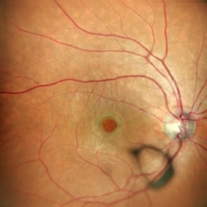
Color fundus photograph of a patient with macular hole along with surrounding cuff of fluid. A Weiss ring can be seen anteriorly in the vitreous.
Photographer: Gayathri Mohan, Retina Foundation
Imaging device: Mirante, Nidek
Condition/keywords: macular hole
-
 Bidding Adieu to Attachments: Weiss Ring
Jan 7 2022 by Gayathri Mohan
Colour fundus photograph showing a Weiss ring following PVD.
Photographer: Dr. GAYATHRI MOHAN
Imaging device: Canon
Condition/keywords: PVD induction, Weiss ring
-
 Evolving Weiss Ring
Sep 11 2022 by Michael B Green, MD, MBA
Fundus photograph of a 62-year-old female with an evolving Weiss-ring in the process of separating from the optic disc.
Condition/keywords: posterior vitreous detachment, PVD, Weiss ring
-
 Peripapillary Glial Proliferation
Oct 18 2012 by Suber S. Huang, MD, MBA, FASRS
61-year-old woman with peripapillary gilal proliferation
Photographer: Stacie Hrvatin
Condition/keywords: glial proliferation, posterior vitreous detachment, Weiss ring
-
 Peripapillary Glial Proliferation
Oct 18 2012 by Suber S. Huang, MD, MBA, FASRS
61-year-old woman with peripapillary gilal proliferation
Photographer: Stacie Hrvatin
Condition/keywords: glial proliferation, posterior vitreous detachment, Weiss ring
-
 Peripapillary Glial Proliferation
Oct 18 2012 by Suber S. Huang, MD, MBA, FASRS
61-year-old woman with peripapillary gilal proliferation
Photographer: Stacie Hrvatin
Condition/keywords: glial proliferation, posterior vitreous detachment, Weiss ring
-
 Peripapillary Glial Proliferation
Oct 18 2012 by Suber S. Huang, MD, MBA, FASRS
61-year-old woman with peripapillary gilal proliferation
Photographer: Stacie Hrvatin
Condition/keywords: glial proliferation, posterior vitreous detachment, Weiss ring
-
 PVD Induction in Progress
Jan 10 2022 by Manish Nagpal, MD, FRCS (UK), FASRS
Intraoperative image of PVD induction with triamcinolone staining clearly showing the Weiss ring, which is being lifted with suction from the cutter.
Photographer: Manish Nagpal, Retina Foundation, Ahmedabad, india
Imaging device: Sony PMW -10 MD surgical camera
Condition/keywords: PVD induction, triamcinolone, Weiss ring
-
 Rhegmatogenous Retinal Detachment, Weiss Ring, Macula Off
Aug 18 2020 by Carlos W Arzabe, MD
Rhegmatogenous retinal detachment, Weiss Ring, macula off.
Imaging device: Clarus 700
Condition/keywords: macula, Weiss ring
-
 Toxoplasmosis Slide 2
Oct 22 2012 by Ronald C. Gentile, MD
Above the optic nerve there was evidence of a Weiss ring. Not unusual since Toxoplasmosis chorioretinitis is a cause of a premature posterior vitreous detachment.
Photographer: The New York Eye & Ear Infirmary Department of Medical Imaging
Condition/keywords: toxoplasmosis
-
 Weiss Ring, Macula Off
Aug 18 2020 by Carlos W Arzabe, MD
Weiss ring, macula off
Imaging device: Calrus 700
Condition/keywords: macula, Weiss ring
-
 Weiss Ring
Jan 9 2019 by John S. King, MD
77-year-old white male with ERM and PVD OD; sheet of vitreous with weiss ring in the nasal mid-vitreous cavity.
Photographer: Macey Highfill, RN
Imaging device: Topcon 50
Condition/keywords: posterior vitreous detachment, Weiss ring
-
 Weiss Ring
Sep 17 2015 by Jason S. Calhoun
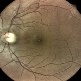
Patient with no visual complaints. Weiss ring visible off the optic dis in both eyes.
Photographer: Jason Calhoun, Mayo Clinic, Department of Ophthalmology
Imaging device: TOPCON-TRC50EX
Condition/keywords: Weiss ring
-
 Weiss Ring
Sep 17 2015 by Jason S. Calhoun
Patient with no visual complaints. Weiss ring visible off the optic dis in both eyes.
Photographer: Jason Calhoun, Mayo Clinic, Department of Ophthalmology
Imaging device: TOPCON-TRC50EX
Condition/keywords: Weiss ring
-
 Weiss Ring
Oct 22 2019 by Jessica Norkus
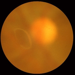
Fundus photo taken on TopCon TRC 50Dx camera of a 60-year-old patient who has experienced an acute PVD. Chief complaint of "large floater" OS prompted exam. Physician noted a significant Weiss Ring and requested fundus color photo for documentation.
Photographer: Jessica Norkus, COA (Retina Specialists of Michigan)
Imaging device: TopCon TRC 50Dx
Condition/keywords: color fundus photograph, color photo, fundus photograph, nerve, optic disc, posterior vitreous detachment, retina, vitreous floaters, Weiss ring
-
 Weiss Ring
Apr 22 2024 by SHIVANG CHAURASIA
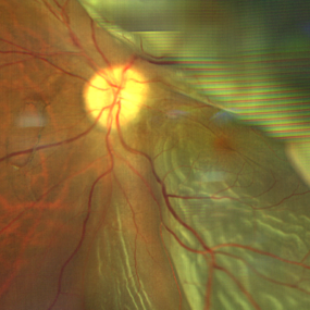
Fundus photograph of a 68-year-old female with complaints of floaters.
Photographer: Dr SHIVANG CHAURASIA, GSVM MEDICAL COLLEGE, KANPUR, UTTAR PRADESH, INDIA
Imaging device: SMARTPHONE FUNDOSCOPY- IPHONE12
Condition/keywords: posterior vitreous detachment, Weiss ring
-
 Weiss Ring
Apr 29 2025 by Gustavo Uriel Fonseca Aguirre
This B-mode axial ultrasound scan demonstrates the Weiss ring, visualized as a circular hyperechoic structure in the vitreous cavity, representing the detached posterior vitreous face with the optic disc insertion site. The ring shows mild mobility on dynamic assessment without retinal traction.
Photographer: Gustavo U. Fonseca Aguirre, Hospital Conde de Valenciana, Ciudad de México
Condition/keywords: Weiss ring
-
 Weiss Ring
Jan 21 2025 by Kimberly Wakester
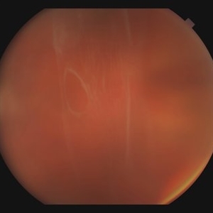
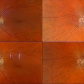
Fundus photographs of a 70-year-old woman with a PVD with Weiss ring present in the left eye. Doing sweeps of the left eye shows how changing the patient's gaze can reposition the Weiss ring in the patient's eye.
Photographer: Kimberly Wakester, COA
Imaging device: Optos California
Condition/keywords: PVD, Weiss ring
-
 Weiss Ring
Jul 13 2013 by Jason S. Calhoun
Weiss ring floater in the right eye.
Photographer: Jason S. Calhoun, Department of Ophthalmology, Mayo Clinic Jacksonville, Florida
Imaging device: TOPCON TRC 50-EX
Condition/keywords: Weiss ring
-
 Weiss Ring
Jan 15 2019 by Olivia Rainey
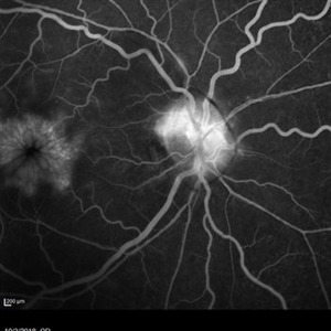
Fluorescein angiogram of a 55-year-old female with a Weiss ring affecting her right eye. Patient was diagnosed with sarcoidosis. She has cystoid macular edema secondary to panuveitis.
Photographer: Olivia Rainey
Imaging device: Heidelberg Spectralis
Condition/keywords: 30 degrees, cystoid macular edema (CME), fluorescein angiogram (FA), fluorescein leakage, Heidelburg Spectralis, optic nerve, sarcoidosis, uveitis, Weiss ring
-
 Weiss Ring
Apr 8 2019 by Gary R. Cook, MD, FACS
Weiss ring ahead of optic disc following a recent posterior vitreous detachment (PVD)
Imaging device: Topcon VT-50
Condition/keywords: posterior vitreous detachment, Weiss ring
-
---thumb.JPG/image-square;max$300,300.ImageHandler) Weiss Ring (Floater)
Weiss Ring (Floater)
Jul 10 2013 by Jason S. Calhoun
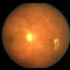
Patient comes in complaining of a floater towards the nasal aspect of her vision. Fundus photograph with anterior shot, shows a weiss ring pulled off from the optic nerve.
Photographer: Jason S. Calhoun, Department of Ophthalmology, Mayo Clinic Jacksonville, Florida
Condition/keywords: floaters, Weiss ring
 Loading…