Search results (43 results)
-
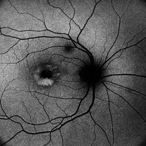
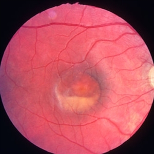
 Best Vitelliform Macular Dystrophy
Jan 14 2018 by Koushik Tripathy, MBBS, MD
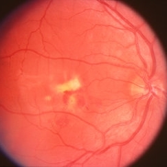
Typical subretinal egg yolk appearance of Best disease. Arden ratio in electrooculogram was reduced in either eye.
Condition/keywords: Best disease, vitelliform macular dystrophy
-
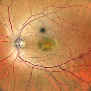
 Adult Onset Vitelliform Macular Dystrophy
Sep 29 2024 by Tejaswita Verma
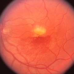
Autofluorescence image of the RE of a 62 year-old hypertensive female with 6/12 vision showing hyperautofluorescence . The patient was asked to review every few months to check for development of secondary CNVM
Photographer: DR. TEJASWITA VERMA
Imaging device: MIRANTE
Condition/keywords: Adult vitelliform macular dystrophy
-
---thumb.jpg/image-square;max$300,300.ImageHandler) Adult Vitelliform Dystrophy
Adult Vitelliform Dystrophy
Feb 13 2013 by From the Collections of Thomas M. Aaberg, MD and Thomas M. Aaberg Jr., MD
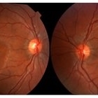
Right eye.
Condition/keywords: vitelliform macular dystrophy
-
---thumb.jpg/image-square;max$300,300.ImageHandler) Adult Vitelliform Dystrophy
Adult Vitelliform Dystrophy
Feb 13 2013 by From the Collections of Thomas M. Aaberg, MD and Thomas M. Aaberg Jr., MD
Left eye
Condition/keywords: left eye, vitelliform macular dystrophy
-
---thumb.jpg/image-square;max$300,300.ImageHandler) Adult Vitelliform Dystrophy
Adult Vitelliform Dystrophy
Feb 13 2013 by From the Collections of Thomas M. Aaberg, MD and Thomas M. Aaberg Jr., MD
Right eye.
Condition/keywords: vitelliform macular dystrophy
-
---thumb.jpg/image-square;max$300,300.ImageHandler) Adult Vitelliform Dystrophy
Adult Vitelliform Dystrophy
Feb 13 2013 by From the Collections of Thomas M. Aaberg, MD and Thomas M. Aaberg Jr., MD
Right eye.
Condition/keywords: vitelliform macular dystrophy
-
---thumb.jpg/image-square;max$300,300.ImageHandler) Adult Vitelliform Dystrophy
Adult Vitelliform Dystrophy
Mar 29 2013 by Henry J. Kaplan, MD
Fundus photograph of the same patient, left eye #2 notice the multiple vitelliform lesions.
Condition/keywords: vitelliform macular dystrophy
-
---thumb.jpg/image-square;max$300,300.ImageHandler) Adult Vitelliform Dystrophy
Adult Vitelliform Dystrophy
Apr 1 2013 by Henry J. Kaplan, MD
Fundus photograph of a middle aged patient with mild decreased vision and bilateral macular vitelliform lesion #1.
Condition/keywords: adult vitelliform dystrophy, vitelliform lesion, vitelliform macular dystrophy
-
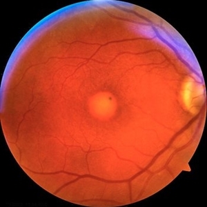
 Adult Vitelliform Macular Dystrophy
Jun 25 2024 by Tejaswita Verma
Left eye fundus photograph of an elderly 62 year old hypertensive female showing elevated lesion at macula s/o adult vitelliform macular dystrophy, misdiagnosed as long standing CSR elsewhere.
Photographer: DR. TEJASWITA VERMA
Imaging device: MIRANTE
Condition/keywords: Adult vitelliform macular dystrophy
-
 Adult Vitelliform Macular Dystrophy
Jun 25 2024 by Tejaswita Verma
Right eye Fundus photo of an elderly 62 year old female with 6/12 vision showing elevated lesion at macula with suggestive of adult vitelliform macular dystrophy, misdiagnosed as long standing CSR elsewhere.
Photographer: DR. TEJASWITA VERMA
Imaging device: MIRANTE
Condition/keywords: Adult-onset vitelliform dystrophy
-
 Adult-onset foveomacular vitelliform dystrophy
May 26 2022 by Rinat Sutiushev
Woman born in 1946. Concerns about decreased vision in the right eye, distortions when reading. The ocular fundus of both eyes shows round yellowish deposits (vitelliform material deposits) in the fovea.
Photographer: Rinat Sutiushev
Condition/keywords: adult vitelliform dystrophy, vitelliform lesion, vitelliform macular dystrophy
-
 Adult-onset foveomacular vitelliform dystrophy
May 26 2022 by Rinat Sutiushev
Woman born in 1946. Concerns about decreased vision in right eye, distortions when reading. The ocular fundus of both eyes shows round yellowish deposits (vitelliform material deposits) in the fovea. When autofluorescence photography is performed, hyperautofluorescence is detected.
Photographer: Rinat Sutiushev
Condition/keywords: adult vitelliform dystrophy, vitelliform lesion, vitelliform macular dystrophy
-
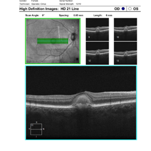 Adult-onset foveomacular vitelliform dystrophy
May 26 2022 by Rinat Sutiushev
Patient born in 1946. Concerns about decreased vision in right eye, distortions when reading. The ocular fundus of both eyes shows round yellowish deposits (vitelliform material deposits) in the fovea. Autofluorescence photography reveals hyperautofluorescence. OCT demonstrates the presence of vitelliform material under the sensitive retina and over the retinal pigment epithelium.
Photographer: Rinat Sutiushev
Condition/keywords: adult vitelliform dystrophy, vitelliform lesion, vitelliform macular dystrophy
-
 Best Disease
Apr 8 2019 by Gary R. Cook, MD, FACS
Right eye of patient with Best disease with active CNV OD; V.A. = 20/30.
Condition/keywords: Best disease, choroidal neovascularization (CNV), vitelliform macular dystrophy
-
 Best Disease
Apr 8 2019 by Gary R. Cook, MD, FACS
Left eye of the patient with Best disease and an active CNV OD showing resolving hemorrhage from prior CNV OS.
Condition/keywords: Best disease, retinal hemorrhage, vitelliform macular dystrophy
-
 Best Disease
Jul 5 2014 by John S. King, MD
Late 30s.
Photographer: URMC
Condition/keywords: Best disease, vitelliform macular dystrophy
-
 Best Disease
Jul 5 2014 by John S. King, MD
30s.
Photographer: URMC
Condition/keywords: Best disease, vitelliform macular dystrophy
-
 Best Disease
Jul 5 2014 by John S. King, MD
20s.
Photographer: URMC
Condition/keywords: Best disease, vitelliform macular dystrophy
-
 Best Disease
Jul 5 2014 by John S. King, MD
Teens.
Photographer: URMC
Condition/keywords: Best disease, vitelliform macular dystrophy
-
 Best Disease
Oct 12 2012 by Gregg T. Kokame, MD, MMM, FASRS
Best disease
Photographer: Jaclyn Pisano, Retina Consultants of Hawaii
Imaging device: Zeiss FF-450 plus
Condition/keywords: Best disease, vitelliform macular dystrophy
-
 Best Disease
Apr 24 2024 by Marcelo Zas, MD PhD
Best vitelliform macular dystrophy (BVMD) or Best disease. Is the most common autosomal dominant macular dystrophy. It involves the retinal pigment epithelium (RPE), and leads to a characteristic bilateral yellow “egg-yolk” appearance of the macula as you can see in this image. Essentially, BVMD is considered to have 6 clinical stages: Previtelliform, Vitelliform, Pseudohypopyon, Vitelleruptive, Atrophic and Choroidal neovascularization. As the disease progresses, patients may experience a slow, bilateral decrease in visual acuity, central scotoma, or metamorphopsia. With secondary CNV, visual decline can be rapid, however.
Photographer: Luciano Scorsetti MD
Condition/keywords: Macular Dystrophy
-
 Best Disease
May 1 2018 by Mitzy E Torres Soriano, MD
Fundus photographs of an 5-year-old boy with best vitelliform macular dystrophy and family history.
Photographer: Luciana García,MD
Condition/keywords: Best disease, vitelliform macular dystrophy
-
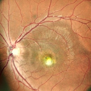
 Best disease left eye
Jan 11 2013 by Alex P. Hunyor, MD
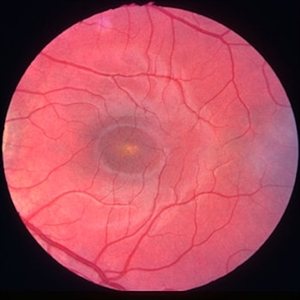
Best's vitelliform macular dystrophy, left eye - early vitelliform stage.
Condition/keywords: Best disease
-
 Best disease, right eye
Jan 11 2013 by Alex P. Hunyor, MD
Best's vitelliform macular dystrophy, right eye - vitelliruptive stage.
Condition/keywords: Best disease
-
 Best Vitelliform Macular Dystrophy
Jun 26 2022 by Vaidehi Sathaye
8 yr old male child with Best Vitelliform Macular Dystrophy
Photographer: Dr. Vaidehi Sathaye
Imaging device: Mirante
Condition/keywords: Best vitelliform macular dystrophy (BVMD)
 Loading…