 Initializing download.
-
 By Rinat Sutiushev
- Uploaded on May 26, 2022.
- Last modified by Joshua Friedman on May 31, 2022.
- Rating
- Appears in
- Retina
- Condition/keywords
- vitelliform macular dystrophy, adult vitelliform dystrophy, vitelliform lesion
- Photographer
- Rinat Sutiushev
- Imaging device
- Optical coherence tomography system
- Description
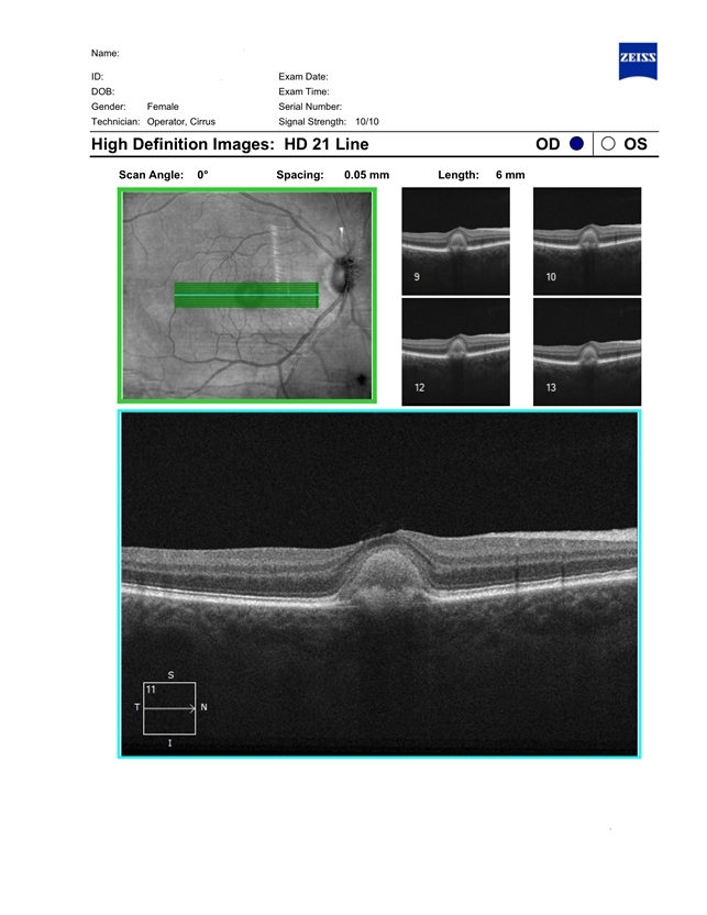
- Patient born in 1946. Concerns about decreased vision in right eye, distortions when reading. The ocular fundus of both eyes shows round yellowish deposits (vitelliform material deposits) in the fovea. Autofluorescence photography reveals hyperautofluorescence. OCT demonstrates the presence of vitelliform material under the sensitive retina and over the retinal pigment epithelium.


---thumb.jpg/image-square;max$79,0.ImageHandler "Multiple Vitellitorn Retinal 2")
---thumb.jpg/image-square;max$79,0.ImageHandler "Multiple Vitelliforn Retinal 3")
---thumb.jpg/image-square;max$79,0.ImageHandler "Multiple Vitelliforn Retinal 7")
---thumb.jpg/image-square;max$79,0.ImageHandler "Multiple Vitelliforn Retinal 6")
---thumb.jpg/image-square;max$79,0.ImageHandler "Multiple Vitelliforn Retinal 5")
---thumb.jpg/image-square;max$79,0.ImageHandler "Multiple Vitelliforn Retinal 4")
---thumb.jpg/image-square;max$79,0.ImageHandler "Adult Vitelliform Dystrophy")