Search results (73 results)
-
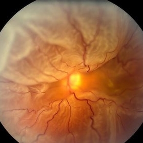
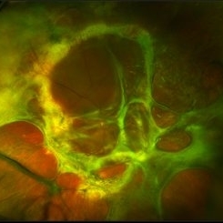
 Total Rhegmatogenous Retinal Detachment With Severe PVR
May 27 2015 by Darin R. Goldman, MD
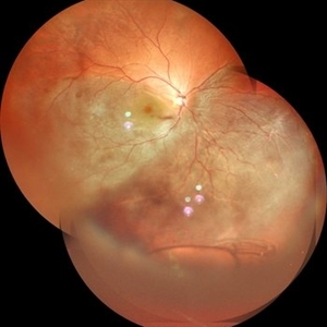
63-year-old pseudophakic male with hand motion vision in the left eye due to a total retinal detachment with severe proliferative vitreoretinopathy.
Condition/keywords: proliferative vitreoretinopathy (PVR), retinal tear
-
 Giant Retinal Tear
Feb 20 2024 by Soobien Lee
Optos color fundus photograph of a 40-year-old caucasian male who is a UFC fighter with a total retinal detachment in his right eye secondary to a giant retinal tear from 10 o'clock to 2 o'clock.
Photographer: Trinity Wolf, Elman Retina Group
Imaging device: Optos Ultra-Widefield Imaging
Condition/keywords: giant retinal tear, optos, Retinal Detachment, Retinal tear with detachment, trauma
-
 360 Degree Retinal Detachment
Jun 29 2013 by Jason S. Calhoun
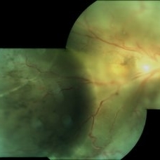
Total retinal detachment in the left eye.
Photographer: Jason S. Calhoun, Mayo Clinic Jacksonville, Florida
Imaging device: TOPCON TRC 50-EX
-
 Horseshoe Retinal Break
Apr 3 2018 by Wesam Safwat
Fundus photograph of an 40-year-old woman with lower temporal horseshoe retinal tear associated with lower sub total retinal detachment not involving macula.
Photographer: Wesam Safwat, Elferdaws eye hospital , Zagazig, Egypt.
Imaging device: Topcon
-
 Macrocyst in the Fovea
Feb 2 2021 by Peter J Belin, MD
36-year-old male with a white cataract and a chronic total retinal detachment for 1 year presented with a recurrent PVR detachment after primary repair 2 weeks prior. This OCT- EDI demonstrates a large retinal cyst through the fovea.
Photographer: Holly Cheshier, CRA, OCT-C, COT
Imaging device: Heidelberg Spectralis
Condition/keywords: chronic retinal detachment, proliferative vitreoretinopathy (PVR), retinal cyst, retinal macrocyst
-
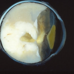 Retinoblastoma
Oct 9 2019 by McGill University Health Centre
Enucleated eye of a pediatric patient showing a total retinal detachment and a large exophytic retinoblastoma.
Photographer: Miguel N. Burnier, McGill University Health Center-McGill University Ocular Pathology & Translational Research Laboratory
Condition/keywords: exophytic, gross pathology, retinoblastoma
-
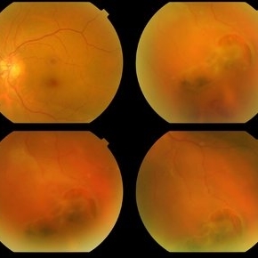
 Total Retinal Detachment With PVR Changes
Nov 5 2018 by awaneesh m upadhyay, MBBS, DNB
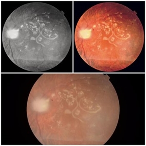
Fundus images of a male pseudophakic patient with total rhegmatogenous retinal detachment and PVR changes.
Photographer: Hiteshwar Saikia
Condition/keywords: proliferative vitreoretinopathy (PVR)
-
 Traumatic Macular Hole with Retinal Detachment and PVR
Sep 27 2012 by Pauline T Merrill, MD, FASRS
Fundus photo of a 13-year-old boy s/p soccer ball injury 1 month previously. In addition to full-thickness macular hole and total retinal detachment with grade C PVR, note pigment granules visible in vitreous over optic nerve.
Photographer: Karen Parque, Illinois Retina Associates, Chicago, IL
Condition/keywords: proliferative vitreoretinopathy (PVR), traumatic macular hole
-
 Total retinal Detachment multiple holes
Sep 26 2022 by Denica Rodriguez
60 year old Male presented with two week old Macula off Retinal detachment with multiple tears.
Photographer: Denica Rodriguez
Imaging device: Optos California
Condition/keywords: color fundus photograph, color photo, macula-off, optos, pseudocolor, Retinal detachment, retinal holes, retinal tear, Retinal tear with detachment, superior arcade, superior field, superior retina, total retinal detachment
-
 Total Retinal Detachment
May 27 2020 by Jason Griffith
25-year-old male with histroy of blunt force trauma, s/p RD repair with hx scleral buckle/cryo.
Photographer: Hollie Sanders, Tennessee Retina, Nashville, TN
Imaging device: Optos California
-
 PVR Retinal Detachment following Laser Retinopexy Slide 1
Oct 22 2012 by Ronald C. Gentile, MD
Acute onset total retinal detachment with PVR 10 weeks following laser retinopexy.
Photographer: The New York Eye & Ear Infirmary Department of Medical Imaging
Condition/keywords: laser retinopexy, proliferative vitreoretinopathy (PVR)
-
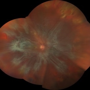 Retinal Detachment
May 1 2019 by Jason Griffith
72-year-old female with advanced total retinal detachment with proliferative vitreoretinopathy.
Photographer: Jason Griffith, Tennessee Retina, Nashville TN
Imaging device: Zeiss Clarus 500
Condition/keywords: proliferative vitreoretinopathy (PVR)
-
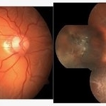
 Acute Retinal Necrosis with Proliferative Vitreoretinopathy and Total Retinal Detachment
Mar 26 2019 by Gary R. Cook, MD, FACS
Same WF patient 9 weeks after initial presentation with Acute Retinal Necrosis now with proliferative vitreoretinopathy and a total combined traction & rhegmatogenous retinal detachment
Imaging device: Topcon VT-50
Condition/keywords: acute retinal necrosis, proliferative vitreoretinopathy (PVR), tractional retinal detachment
-
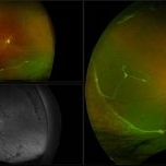 Chronic Retinal Detachment with Proliferative Vitreoretinopathy
Jan 25 2024 by Isaac Agranoff
Widefield fundus photography of a 24 year old male presenting with subtotal retinal detachment with circumferential anterior proliferative vitreoretinopathy. The detachment is bullous inferiorly with atrophic retina and subretinal bands. There are also scattered patches of lattice with atrophic holes and associated detachment in the periphery. Patient presented with flashes for 2 years with worsening vision over the past 6-8 months, measured at 20/100 ph 20/60 OS.
Photographer: Isaac Agranoff, Ashley Rigdon
Imaging device: Optos California
Condition/keywords: atrophic hole, chronic retinal detachment, lattice degeneration, proliferative vitreoretinopathy (PVR), subretinal bands
-
 Chronic Total Retinal Detachment
Oct 12 2012 by Jeffrey G. Gross, MD, FASRS
Chronic, total RD, with shifting inferior subretinal fluid.
Condition/keywords: chronic, inferior subretinal fluid
-
 Chronic Total Retinal Detachment
Oct 8 2019 by Olivia Rainey
Ultra-wide field pseudocolor image of a 57-year-old male with a chronic total retinal detachment affecting his right eye. Patient presented with klebsiella endophthalmitis in UK, and was in medically induced coma with tracheostomy. He awoke after sedation with loss of vision in both eyes, later developing a retinal detachment in both eyes. He had his first repair with gas then with silicone oil. Although he is developing band K and corneal decompensation due to oil in the AC, he has a chronic cicatricial retinal detachment with subretinal and preretinal PVR with large stretch breaks. His vision may worsen with surgery and his eye is hypotenous and if repair is possible, oil will need to be replaced and thus may still migrate to the AC thus would defer intervention until absolutely necessary given risks of vision loss in his only seeing eye. Given this is his only eye, observation is recommended at this time.
Photographer: Olivia Rainey and Amber Poss
Imaging device: Optos
Condition/keywords: chronic retinal detachment, klebsiella endopthalmitis, laser scarring, Optos, proliferative vitreoretinopathy (PVR), retinectomy, silicone oil
-
 Dislocated IOL in Vitreous with RD
May 11 2020 by Gayathri Mohan
Color fundus photo montage of a patient showing a dislocated posterior chamber intraocular lens in the vitreous cavity inferiorly along with a sub total retinal detachment.
Photographer: Gayathri Mohan, Retina Foundation
Imaging device: Mirante, Nidek
Condition/keywords: dislocated posterior chamber intraocular lens (PCIOL), montage
-
 Emulsified Silicone Oil in Macular Hole
Jun 7 2024 by Vaidehi Jethwa
Fundus photograph of 72 year old man was having a/h/o Left Eye trauma by a cow horn, 8 years Ago, and developed Left Eye total Retinal detachment and was operated for Left Eye vitrectomy with FAX, SOI, Endolaser on 11 /04/2015 and was advised Left Eye silicone Oil removal.
Photographer: Dr. Vaidehi Jethwa, M and J institute of Ophthalmology, Ahmedabad, Gujarat.
Imaging device: Zeiss Visucam lite
Condition/keywords: macular hole, silicon oil emulsification in vitreous cavity
-
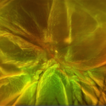
 Familial Exudative Vitreoretinopathy
Jan 11 2018 by S. Natarajan, MD, FASRS, FRCS (GLASGOW) , FICO, D.Sc, FELA
Fundus photograph of 25-year-old female with Familial Exudative Vitreoretinopathy ; right eye showing total retinal detachment with proliferative vitreoretinopathy.
Photographer: miss Ashwini borde
Imaging device: Carl Zeiss 450 plus IR
Condition/keywords: familial exudative vitreoretinopathy (FEVR)
-
 Funnel Retinal Detachment With Proliferative Vitreoretinopathy
Oct 2 2013 by Jerald A. Bovino, MD
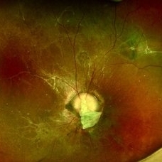
There is a total retinal detachment. The proliferative vitreoretinopathy has caused the retina to assume a funnel shape.
Condition/keywords: funnel, proliferative vitreoretinopathy (PVR), re-attached retinal detachment (RRD)
-
 Funnel Retinal Detachment With Proliferative Vitreoretinopathy
Oct 2 2013 by Jerald A. Bovino, MD
There is a total retinal detachment. The proliferative vitreoretinopathy has caused the retina to assume a funnel shape.
Condition/keywords: funnel, proliferative vitreoretinopathy (PVR), re-attached retinal detachment (RRD)
-
 Hypotony, Thickend Choroid, Total Retinal Detachment, Foreshortened Globe
Dec 10 2012 by Yale L. Fisher, MD
Foreshortening in a phtisical eye with a thickened choroid.
Condition/keywords: video
-
 Morning Glory Anomaly With Retinal Detachment Managed With Amniotic Membrane Graft
Oct 15 2024 by Hemanth Murthy, MBBS, MD, FASRS
10 year-old boy presented with noticed blurring of vision. He had total retinal detachment with complicated cataract. He underwent lensectomy with 240 band and vitrectomy with silicone oil. The retina failed to settle due to minute breaks in the inferior part of the disc. Repeat surgery with AMG was done to cover the inferior part of disc. The retina settled under silicone oil. Silicone oil was removed and he is presently undergoing amblyopia treatment. Vision is 2/60 with +14.5 diopter lens.
Photographer: Mr Veda Vyas
Condition/keywords: amniotic membrane graft, Morning Glory Anomaly
-
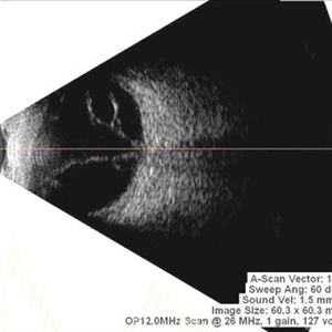 Multiple Retinal Cysts Associated With Chronic Retinal Detachment
Sep 24 2018 by samarth mishra
Patient presented with a diminution of vision in left eye since few months. On B-scan ultrasonography multiple retinal cysts with a total retinal detachment were noted.
Photographer: Aditya Birla Sankara Nethralaya, West Bengal , Kolkata , India
Condition/keywords: B scan ultrasound, chronic retinal detachment, intraretinal cyst, retinal cyst
-
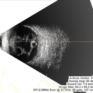 Multiple Retinal Cysts Associated With Chronic Retinal Detachment
Sep 24 2018 by samarth mishra
Patient presented with a diminution of vision in left eye since few months. On B-scan ultrasonography multiple retinal cysts with a total retinal detachment were noted.
Photographer: Aditya Birla Sankara Nethralaya, West Bengal , Kolkata , India
Condition/keywords: B scan ultrasound, chronic retinal detachment, intraretinal cyst, retinal cyst
 Loading…