Search results (29 results)
-
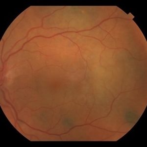 BDUMP
BDUMP
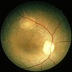
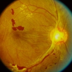
Dec 11 2018 by John S. King, MD
67-year-old white female with normal vision four months ago, consulted for dry AMD. She reported that vision in the left eye had worsened over the last two months and had progressively gotten worse. Denied history of cancer, or her primary eye doctor ever mentioning choroidal nevi. Va cc was 20/30 OD and 20/100 OS. No RAPD. IOP 9-10 OU. Anterior segment had some stellate like pigmented dusting of the endothlium, a/c was quiet, 2+NSC OU. Vitreous quiet; multiple, flat, pigmented choroidal lesions varying in size was seen the in fundus. Area in the temporal macula extending up to the superior arcade in the left eye that was suspicious for a mass; it did have a "giraffe like" pattern on one of the early FA pics; the OCT in this area showed thickening of the choroid without a definite mass lesion, and overlying thickening of the RPE, or exudative like scar, with SRF directly above. Consulted with Dr. Matt Wilson, who confirmed diagnosis, and had patient evaluated by oncology, who diagnosed non-small cell lung cancer.
Photographer: Stacey Coleman
Imaging device: Topcon
Condition/keywords: bilateral diffuse uveal melanocytic proliferation (BDUMP)
-
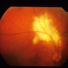
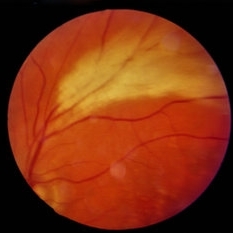
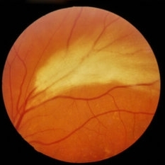
 Total retinal Detachment multiple holes
Sep 26 2022 by Denica Rodriguez
60 year old Male presented with two week old Macula off Retinal detachment with multiple tears.
Photographer: Denica Rodriguez
Imaging device: Optos California
Condition/keywords: color fundus photograph, color photo, macula-off, optos, pseudocolor, Retinal detachment, retinal holes, retinal tear, Retinal tear with detachment, superior arcade, superior field, superior retina, total retinal detachment
-
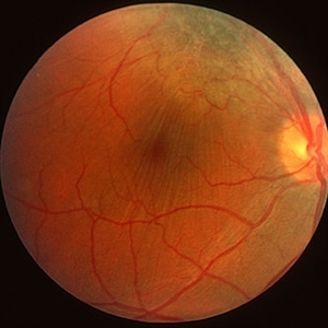
 Retinal Angiomas In VHL
Dec 24 2012 by Roy D. Brod, MD
Fundus photograph of 16 year old male with recent diagnosis of Von Hippel-Lindau disease showing typical appearance of a retinal angioma in superior mid periphery OD. Note unrelated choroidal nevus above superior arcade.
Photographer: Julia Walker
Condition/keywords: hemangioma, Von Hippel-Lindau
-
 AIDS - Toxoplasmosis
Jun 4 2014 by Henry J. Kaplan, MD
Fundus photograph of a patient with AIDS who has developed toxoplasma retinochoroiditis; large yellow patch of retinitis along the superior arcade fading the vasculature with feathery edges. #1
Condition/keywords: AIDS, toxoplasmosis
-
 Angiomatosis Retinae
Jul 22 2020 by MOHIT GUPTA
39-year-old female presented with retinal angiomas along superior arcade with macular exudation.
Photographer: Dr Mohit Gupta, Prakash Netra Kendr, Lucknow, India
Imaging device: Heidelberg spectralis Fundus camera
Condition/keywords: angiomatosis retinae
-
 BDUMP
BDUMP
Dec 11 2018 by John S. King, MD
67-year-old white female with normal vision four months ago, consulted for dry AMD. She reported that vision in the left eye had worsened over the last two months and had progressively gotten worse. Denied history of cancer, or her primary eye doctor ever mentioning choroidal nevi. Va cc was 20/30 OD and 20/100 OS. No RAPD. IOP 9-10 OU. Anterior segment had some stellate like pigmented dusting of the endothlium, a/c was quiet, 2+NSC OU. Vitreous quiet; multiple, flat, pigmented choroidal lesions varying in size was seen the in fundus. Area in the temporal macula extending up to the superior arcade in the left eye that was suspicious for a mass; it did have a "giraffe like" pattern on one of the early FA pics; the OCT in this area showed thickening of the choroid without a definite mass lesion, and overlying thickening of the RPE, or exudative like scar, with SRF directly above. Sent patient to Dr. Matt Wilson, who confirmed BDUMP, and had patient sent to oncology to find a possible primary lesion. Mass seen on CT chest; biopsy revealed non-small cell lung cancer, and is getting chemo/radio treatment. Ocular findings have not progressed over the last few months.
Photographer: Stacey Coleman
Imaging device: Topcon 50
Condition/keywords: bilateral diffuse uveal melanocytic proliferation (BDUMP)
-
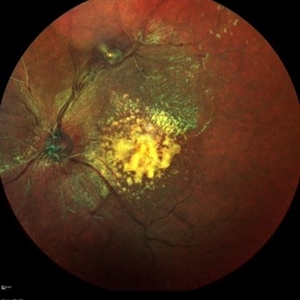
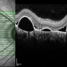 Chronic Central serous chorioretinopathy
Nov 10 2022 by T. P . VIGNESH, MBBS,MS
SD-OCT of a 45 year old man , revealing multiple serous retinal pigment epithelial detachments (PED) just inferior to superior arcade .
Photographer: Priyanka
Imaging device: Heidelberg Spectralis
Condition/keywords: chronic central serous chorioretinopathy (CSCR)
-
---thumb.jpg/image-square;max$300,300.ImageHandler) CMV Retinitis in a Patient with the Diagnosis of AIDS
CMV Retinitis in a Patient with the Diagnosis of AIDS
Feb 27 2013 by Henry J. Kaplan, MD
CMV retinitis, left eye: classic form in AIDS patient. Hemorrhagic retinitis mainly in the superior arcade.
Condition/keywords: AIDS
-
 Combined Hamartoma of Retina and RPE
Mar 29 2013 by Henry J. Kaplan, MD
Hamartoma visible as a grreen lesion on superior arcade with ERM formation and dragging of the macula.
Condition/keywords: combined hamartoma
-
 ERMageddon - Wrinkle in the Space-time Fabric of Macula
Oct 29 2025 by SHRADDHA RAJ SHRIVASTAVA
38 year old female with Epiretinal Membrane (ERM) over macula, post laser barrage for multiple symptomatic Horse-shoe Tears (HSTs) and Lattice Degenerations. Posterior pole revealed tilted disc with peripapillary atrophy. There is thick opaque epiretinal membrane obscuring the underlying superior arcade vessels and causing foveal ectopia with distortion of perimacular vasculature. Patient was planned for Right Eye pars plana vitrectomy for ERM peeling.
Photographer: Dr. Shraddha Raj Shrivastava
Imaging device: Nidek Mirante SLO/OCT (Confocal scanning/Spectral domain OCT
Condition/keywords: ectopic fovea, epiretinal membrane (ERM), ERM, horseshoe tear, vitreomacular traction (VMT)
-
 ERMageddon - Wrinkle in the Space-time Fabric of Macula
Oct 29 2025 by SHRADDHA RAJ SHRIVASTAVA
38 year old female with Epiretinal Membrane (ERM) over macula, post laser barrage for multiple symptomatic Horse-shoe Tears (HSTs) and Lattice Degenerations (seen on wide-field image). Posterior pole revealed tilted disc with peripapillary atrophy. There is thick opaque epiretinal membrane obscuring the underlying superior arcade vessels and causing foveal ectopia with distortion of perimacular vasculature. Patient was planned for Right Eye pars plana vitrectomy for ERM peeling.
Photographer: Dr. Shraddha Raj Shrivastava
Imaging device: Nidek Mirante SLO/OCT (Confocal scanning/Spectral domain OCT
Condition/keywords: BARRAGE LASER, ectopic fovea, epiretinal membrane (ERM), horseshoe tear, lattice degeneration, vitreomacular traction (VMT)
-
 Hemi Central Retinal Artery Occlusion
Apr 17 2024 by Akansha Sharma
Early phase fluorecein angiogram of a 30 year old female with perfusion lacking along the vessels of the superior arcade.
Photographer: Dr. Akansha Sharma, Bharati Eye Hospital
Condition/keywords: central retinal artery occlusion (CRAO), CRAO, Hemi-Central Retinal Artery Occlusion (CRAO)
-
 Hemi Vein Occlusion, Fluorescein Angiogram, Montage
Dec 17 2015 by James B. Soque, CRA, OCT-C, COA, FOPS
74-year-old woman, with recurrent superior hemi vein occlusion, montage image of fluorescein angiogram left eye. Currently receiving Lucentis injections OS.
Photographer: James Soque, CRA COA
Imaging device: Topcon RC 50 DX Fundus Camera with MERGE Winstation Software for Fluorescen Angiography
Condition/keywords: montage, occlusion of retinal vein, superior arcade
-
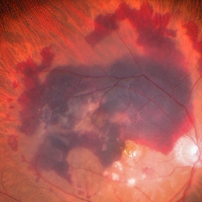 Large Subretinal Bleed in Case of Wet ARMD
Sep 28 2024 by Anjana Mirajkar, MS Ophthalmology
An intra operative image showing large sub retinal hemorrhage involving the macular area and along the superior arcade with exudation at the macular area in case of wet ARMD.
Photographer: Dr. Anjana Mirajkar -Retina Foundation, Ahmedabad
Condition/keywords: polypoidal choroidal vasculopathy (PCV), subretinal hemorrhage, wet age-related macular degeneration (wet AMD)
-
 Light Toxicity
Mar 29 2013 by Henry J. Kaplan, MD
Microscope light toxicity in superior arcade.
Condition/keywords: light toxicity
-
 Macula sparing Superior Rhegmatogenous retinal detachment
Feb 15 2018 by Kushal S Delhiwala, MBBS, MS, FMRF,FICO, FAICO
60- year-old phakic female presenting with sudden onset floaters and curtain like shadow in inferior field of vision in right eye, having undergone scleral buckling surgery in left eye before 2 years. Color fundus photograph montage of right eye showing fresh superior rhegmatogenous retinal detachmnent sparing macula and well above superior arcade.
Photographer: Dr Kushal Delhiwala, Netralaya superspeciality eye hospital ,Ahmedabad
Imaging device: Zeiss Visucam 500
Condition/keywords: macula sparring
-
 Macular PVR. Before and After Scleral Buckle + Vitrectomy
Apr 27 2023 by Jesus Lozano, MD
64 year old woman. Presented to our clinic with PVR related retinal detachment in the left eye. Picture 1 - RRD with a posterior star fold present along the superior arcade. Picture 2 - Postoperative Posterior Scleral Buckle + PPV showing an attached retina with good scleral buckle indentation.
Imaging device: Optos
-
 Myelinated Nerve Fiber Layer
Jan 30 2015 by H. Michael Lambert, MD
Embryonic myelin on superior arcade retina.
Condition/keywords: myelinated nerve fiber layer
-
 Myelinated Nerve Fiber Layer
Jan 30 2015 by H. Michael Lambert, MD
Embryonic myelin on superior arcade retina.
Condition/keywords: myelinated nerve fiber layer
-
 OCT en face of a 360 retinotomy for closed funnel combined retinal detachment
Jan 1 2023 by Malek Yassine, MD
Swept source OCT en face at the silicon oil - Retina Interface shows droplets of SO emulsification around the fovea and at the superior arcade, with some inferior striae corresponding to ERM formation
Imaging device: Topcon Triton DRI-OCT
Condition/keywords: oct en face
-
 Proliferative Diabetic Retinopathy (PDR)
Sep 14 2023 by Ben Serar
Fundus photograph of RE showing traction at the disc, pre retinal haemorrhages and exudates along the superior arcade, and fibrovascular proliferation along the inferior arcade in a case of Proliferative Diabetic Retinopathy (PDR).
Condition/keywords: proliferative diabetic retinopathy (PDR)
-
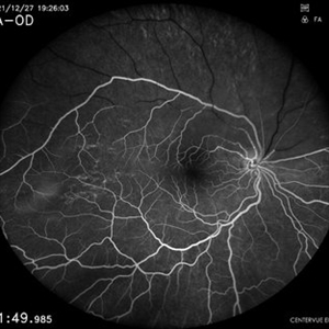
 Proliferative Diabetic Retinopathy - Neovascularization Elsewhere
Nov 11 2013 by Gerardo Garcia-Aguirre, MD
Fluorescein angiogram showing leakage secondary to neovascularization in the superior arcades.
Condition/keywords: neovascularization (NV)
-
 Proliferative Diabetic Retinopathy - Neovascularization Elsewhere
Nov 11 2013 by Gerardo Garcia-Aguirre, MD
Fluorescein angiogram showing leakage secondary to neovascularization in the superior arcades.
Condition/keywords: neovascularization (NV)
-
 Proliferative Diabetic Retinopathy - Neovascularization Elsewhere
Nov 11 2013 by Gerardo Garcia-Aguirre, MD
Fluorescein angiogram showing leakage secondary to neovascularization in the superior arcades.
Condition/keywords: neovascularization (NV)
 Loading…