Search results (282 results)
-
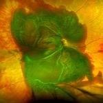
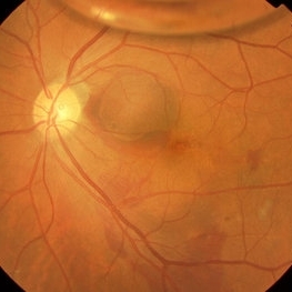
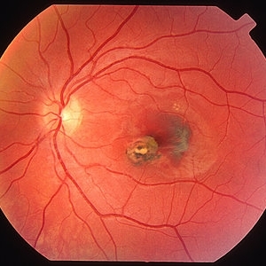
 Massive Subretinal Hemorrhage With Near Total Retina Detachment
Nov 27 2013 by David W. Faber, MD
Fundus photo of an 71-year-old male with massive subretinal hemorrhage. Had been given 6 week Avastin treatments. Was put on coumadin for 6 weeks following knee surgery.
Photographer: Donna Knight, Rocky Mountain Retina Consultants, Salt Lake City, Utah
-
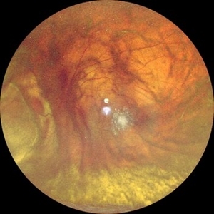
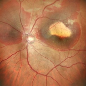
 Choroidal Melanoma
Nov 3 2022 by pedro fernandes souza neto
Transillumination of Enucleation specimen of Choroidal Melanoma: anterior chamber is closed. Total secondary retinal detachment with subretinal serous fluid and some subretinal hemorrhages are present.
Photographer: Eduardo Marback, Federal University of Bahia, Brazil
Condition/keywords: enucleation, melanoma
-
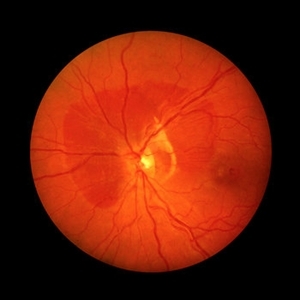
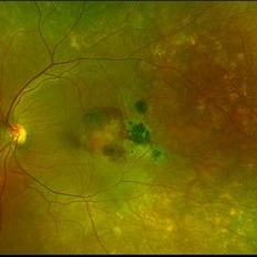
 Lady in a dress
Feb 9 2023 by Shelby Helton
Fluorescein Angiography on a 67-year-old male with significant RPE changes secondary to a severe subretinal hemorrhage that required a vitrectomy with subretinal TPA in 2013.
Photographer: Shelby Helton
Imaging device: Heidelberg Spectralis
Condition/keywords: wet age-related macular degeneration (wet AMD)
-
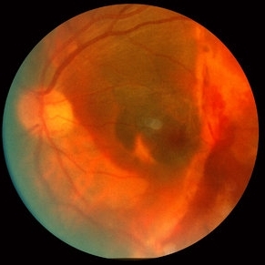
 Subretinal BSS and air
Apr 12 2022 by Nassim Alejandro Abreu Arbaje, MD
67 year old female who presented with complaints of 5 days of decreased vision of her left eye. She underwent PPV + BSS and Air injection in the subretinal space
Photographer: Nassim Abreu, Dr. Elias Santana Hospital
Imaging device: Ngenuity 3D system screenshot
Condition/keywords: subretinal hemorrhage
-
 Massive SRH in Patient on Coumadin Being Treated for Exudative AMD
Sep 30 2019 by John S. King, MD
78-year-old white female using 1mg of warfarin for atrial fibrillation, who had a large PED, Type 1 lesion from AMD. Noticed acute darkening of vision one week after anti-VEGF injection. Has very large SRH, subRPE heme, and corrugated retinal appearance post RPE tear. Vision HM (from 20/100). 20/25 in the fellow eye that has dry AMD.
Photographer: Shelly Blair
Imaging device: Optos CA
Condition/keywords: subretinal hemorrhage, wet age-related macular degeneration (wet AMD)
-
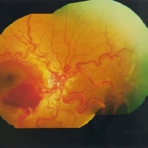
 PEHCR (Peripheral Exudative Hemorrhagic Chorioretinopathy)
May 12 2023 by Niloofar Piri, MD
Ultrawide fundus photograph of the left eye demonstrating extensive peripheral hemorrhagic exudative detachment in a 79 yo Caucasian female with prior history of non-exudative AMD. Recent diagnosis of Acute myeloid leukemia with low platelet count which might have contributed to the above presentatuon. Please note the temporal subretinal hemorrhage as well as RPE atrophy and hyperplasia in the macula.
Photographer: Rocio Bentivegna, MD, Saint Louis University; Jessica Maddox, COA, Saint Louis University
Condition/keywords: peripheral exudative hemorrhagic chorioretinopathy (PEHCR)
-
---thumb.jpg/image-square;max$300,300.ImageHandler) Active Choroidal Neovascularization With Subretinal Hemorrhage
Active Choroidal Neovascularization With Subretinal Hemorrhage
Nov 25 2013 by Maurice F. Rabb
Active choroidal neovascularization with subretinal hemorrhage.
Condition/keywords: choroidal neovascularization (CNV), subretinal hemorrhage
-
 Branch Retinal Vein Occlusion
Oct 17 2012 by Sharon Fekrat, MD FACS FASRS
Fluorescein angiography of an inferior perfused branch retinal vein occlusion in the left eye of a middle aged male with hypertension. The foveal avascular zone is irregular. Subretinal hemorrhage is present.
Photographer: John Reaves, Ophthalmic Photographer, Durham VA Medical Center Eye Clinic Imaging Suite, Durham, NC
Imaging device: Fluorescein Angiography
Condition/keywords: branch retinal vein occlusion (BRVO), subretinal hemorrhage
-
 Choroidal Melanoma Masquerading as Subretinal Hemorrhage With Breakthrough VH
Jan 23 2025 by Tejaswita Verma
A 65 year old diabetic male presented with large nasal retinal mass giving the appearance of organized dehaemoglobinized subretinal hemorrhage with breakthrough vitreous hemorrhage , with 6/6P vision. Enucleation specimen showed histopathology confirmed choroidal melanoma.
Photographer: DR. TEJASWITA VERMA
Imaging device: MIRANTE
-
 Choroidal Nevus with Subretinal Hemorrhage
Jan 29 2015 by Gordon Finnerty
47-year-old woman diagnosed with choroidal nevus with subretinal hemorrhage.
Photographer: Gordon Finnerty
Imaging device: Topcon TRC -50DX
Condition/keywords: choroidal nevus, subretinal hemorrhage
-
 Choroidal Rupture with Subretinal Hemorrhage
Oct 1 2012 by Jeffrey G. Gross, MD, FASRS
Choroidal rupture with subretinal hemorrhage.
Condition/keywords: choroidal rupture, subretinal hemorrhage
-
 Color Montage Picture
Jan 15 2019 by Aniruddha Maiti, MBBS DO DNB FRVS FICO MRCS FACS FASRS FRCOphth
Dilated tortuous vessels with intraretinal and subretinal hemorrhage at fovea.
Photographer: Sangeeta Mohanta
Imaging device: Zeiss FF450 plus IR
Condition/keywords: dilated tortuous vessels, subretinal hemorrhage, Wyburn-Mason
-
 Subfoveal Subretinal Hemorrhage
Oct 15 2012 by Sharon Fekrat, MD FACS FASRS
74-year-old female with subfoveal subretinal hemorrhage due to neovascular AMD.
Photographer: Duke University Eye Center, Durham, NC
Condition/keywords: subretinal hemorrhage
-
 Submacular Hemorrhage After Pneumatic Displacement
Mar 21 2013 by Yusuke Oshima, MD, PhD
Fundus photograph demonstrates an effective displacement of the subretinal hemorrhage from the fovea.
Photographer: Yusuke Takada, Osaka University Graduate School of Medicine
Condition/keywords: submacular hemorrhage
-
 Subretinal Hemorrhage around Disc
Feb 12 2021 by ASRS Staff
Fundus photograph of 38-year-old man with subretinal hemorrhage around the disc.
Imaging device: Nidek Mirante
Condition/keywords: disc, fovea, subretinal hemorrhage
-
 Subretinal Hemorrhage with Chorioretinal Macular Scars
Sep 28 2022 by Denica Rodriguez
Ultra-widefield pseudocolor fundus photograph of a 59 year old female with Subretinal Hemorrhage with Chorioretinal Macular Scars affecting her left eye. The physician presumes the etiology is CNV from adjacent scarring/choroidal rupture. Patient has history of ocular trauma with cricket ball at age 10-12 years old. She suspects that she might have suffered choroidal rupture, which has resulted in secondary CNV and hemorrhage that we are seeing today. She recommends treatment with Eylea sample injection in a series of 4 at a 4-5 week interval. The patient's vision at the time of her appointment was Dcc20/40-2 PHNI.
Photographer: Denica Rodriguez, COA
Imaging device: Optos California
Condition/keywords: antiVEGF therapy, chorioretinal scar, choroidal neovascular membrane (CNVM), fundus photography, left eye, macular scar, Optos, peripheral drusen, pseudocolor, secondary CNV, subretinal hemorrhage, ULTRA WIDE FIELD, ultra-wide field imaging
-
 Surgical Displacement of Subfoveal Subretinal Hemorrhage Using rt-PA, Postop Day One
Oct 15 2012 by Sharon Fekrat, MD FACS FASRS
Fundus photograph of a 76 year old female with neovascular AMD who developed a subfoveal subretinal hemorrhage in the left eye. This photograph is one day postop after 23g vitrectomy, subretinal rt-PA 50ug/0.1cc and C3F8 gas. Note the subretinal hemorrhage and fluid displaced inferiorly. No open retinal break was present.
Photographer: Duke University Eye Center, Durham, NC
-
 Subfoveal Subretinal Hemorrhage
Aug 28 2012 by Sharon Fekrat, MD FACS FASRS
subfoveal subretinal hemorrhage, right eye.
Photographer: Michael P. Kelly, FOPS Director, Duke Eye labs Duke University Eye Center Durham, NC
Imaging device: Zeiss FF40
Condition/keywords: subretinal hemorrhage
-
 Arteriolar Macroaneurysm
Oct 1 2012 by Jeffrey G. Gross, MD, FASRS
Arteriolar macroaneurysm with partially reabsorbed subretinal hemorrhage.
Condition/keywords: arteriolar macroaneurysm, partially reabsorbed subretinal hemorrhage
-
 Ruptured Macroaneurysm
May 22 2019 by Nichole Lewis
FA of a 91-year-old woman with a ruptured macroaneurysm, intraretinal hemorrhage and subretinal hemorrhage. VA 20/400.
Photographer: Nichole Lewis
Condition/keywords: intraretinal hemorrhage, ruptured macroaneurysm, subretinal hemorrhage
-
 Subhyaloid Hemorrhage
Oct 8 2012 by Jeffrey G. Gross, MD, FASRS
Subhyaloid hemorrhage, layered, with surrounding subretinal hemorrhage.
Condition/keywords: subhyaloid hemorrhage, subretinal hemorrhage
-
 Choroidal Hemorrhage, Subretinal Hemorrhage
Dec 18 2017 by Nichole Lewis
Choroidal hemorrhage, Subretinal Hemorrhage, wet macular degeneration,
Photographer: Nichole Lewis
Condition/keywords: choroidal hemorrhage, choroidal neovascularization (CNV), exudative age-related macular degeneration, subretinal hemorrhage, wet age-related macular degeneration (wet AMD)
-
 Choroidal Rupture with Extensive Subretinal Hemorrhage HM
Oct 1 2012 by Jeffrey G. Gross, MD, FASRS
Choroidal rupture with extensive subretinal hemorrhage HM.
Condition/keywords: choroidal rupture, HM, subretinal hemorrhage
-
 Secondary Choroidal Neovascularization Due to Toxoplasmosis
Feb 25 2013 by Henry J. Kaplan, MD
Left eye: secondary choroidal neovascularization and subretinal hemorrhage in a patient with old macular scar of toxoplasma.
Condition/keywords: choroidal neovascularization (CNV), toxoplasmosis, toxoplasmosis chorioretinitis
-
 Subretinal Hemorrhage
Jan 7 2020 by Stacie Neview
Optos fundus photograph of a 74-year-old male with severe subretinal hemorrhage and exudative retinal detachment secondary to peripheral choroidal neovascular membrane.
Photographer: Stacie Neview, Retina Specialists of Michigan, Grand Rapids Michigan, USA
Imaging device: Optos California
Condition/keywords: exudative retinal detachment, subretinal hemorrhage
 Loading…