Search results (274 results)
-
 Central Serous Retinopathy
Mar 19 2024 by Corey Grant
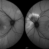
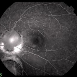
Ultra Wide-Field Fundus Autofluorescence Imaging of a 37 year old female with Central Serous Retinopathy affecting her right eye. Patient Visual Acuity was 20/20 in both eyes. Patient reported black spots in her vision onset three years ago, with associating flashes of light. Patient reports history of cortisone back injections a few years ago and denies Flonase use. The physician stated that there is hyperautofluorescence in the area of gutter of Sub-Retinal Fluid which likely happened from CSR.
Photographer: Corey Grant, OSC
Imaging device: OPTOS CALIFORNIA RGB
Condition/keywords: Central Serous Chorioretinopathy (CSR), central serous retinopathy (CSR), fundus autofluorescence (FAF), Guttering, hyperautofluorescence, inferior retina, OPTOS, Retina, Right Eye, subretinal fluid, ULTRA WIDE FIELD
-
 Idiopathic Uveal Effusion Syndrome
Aug 22 2024 by Jordyn Beckman
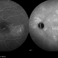
61 year old male with Idiopathic Uveal Effusion Syndrome with starry night appearance on fluorescein. 3 weeks s/p single external drainage retinotomy and 9 weeks of oral pred with recurrent choroidal effusions. Has since returned to surgery for secondary drainage retinotomy; subretinal fluid remain persistent.
Photographer: Jordyn Beckman
Imaging device: Optos California
Condition/keywords: chorioretinitis, Choroidal, exudative detachment, window defect
-
 Amelanotic Choroidal Melanoma
May 18 2020 by McGill University Health Centre
The enucleation specimen in (A) shows an amelanotic, mushroom-shaped tumor arising from the choroid. Microhemorrhages are present within the tumor and also surround the tumor base (arrow). True retinal detachment is present (arrowhead). The subretinal fluid is mixed: clear (1), hemorrhagic (2), and fibrinoid (3).
Condition/keywords: enucleation, mushroom-shaped
-
 Amelanotic Mushroom-Shaped Choroidal Melanoma
May 18 2020 by McGill University Health Centre
This enucleation specimen demonstrates an amelanotic, mushroom-shaped, slightly hemorrhagic tumor near the optic nerve (arrow). True retinal detachment is present, and the retina is folded (arrowhead). The subretinal fluid is hazy (*).
Condition/keywords: amelanotic melanoma, enucleation, mushroom-shaped
-
 Bullous Retinal Detachment
Jul 9 2021 by Anton Orlin, MD
This is a color photograph of a right eye with a superior, bullous, macula-splitting retinal detachment. These features place the patient at a higher risk for macular fold formation postoperatively. To prevent fold formation, a surgeon should attempt for more complete subretinal fluid drainage during repair. This can be done with the use of perfluorocarbon liquid or by making a drainage retinotomy.
Condition/keywords: macular fold
-
 Central Serous Chorioretinopathy
Jan 25 2022 by Olivia Rainey
Widefield fundus autofluorescence of a 60-year-old male with Central Serous Chorioretinopathy affecting both eyes. Chronic history of CSR followed with observation without treatment prior to presenting at our office. The physician noted significant findings on exam and imaging with multifocal areas of inactive and active changes in the right eye and subfoveal subretinal fluid with recent visual decline in the left eye. There are hyper and hypoautofluorescent changes, consistent with CSR.
Photographer: Olivia Rainey, OCT-C, COA
Imaging device: Heidelberg Spectralis
Condition/keywords: 55-degrees, central serous chorioretinopathy (CSCR), central serous retinopathy (CSR), chronic central serous chorioretinopathy (CSCR), fundus autofluorescence (FAF), heidelberg spectralis, left eye
-
 Central Serous Chorioretinopathy
Jan 25 2022 by Olivia Rainey
Late phase widefield fluorescein angiography of a 60-year-old male with Central Serous Chorioretinopathy. Chronic history of CSR followed with observation without treatment prior to presenting at our office. The physician noted subfoveal subretinal fluid with recent visual decline. FA shows multifocal leakage and ICG shows hypercyanescence. OCTA, ICG, and FA consistent with CSR, and without concern for CNVM thus will observe without anti-VEGF at this time. PDT therapy recommended.
Photographer: Olivia Rainey, OCT-C, COA
Imaging device: Heidelberg Spectralis
Condition/keywords: 55-degrees, central serous chorioretinopathy (CSCR), central serous retinopathy (CSR), chronic central serous chorioretinopathy (CSCR), fluorescein angiogram (FA), heidelberg spectralis, indocyanine green (ICG) angiography, left eye
-
 Central Serous Choroidopathy, CSR, with Foci of Leakage
Oct 9 2012 by James B. Soque, CRA, OCT-C, COA, FOPS
50 y/o WM with Central Serous Choroidopathy Left eye. VA OS cc 20/80. Topcon 3D 1000 SD OCT image reveals Sub RPE detachment in several locations, and subretinal fluid blister. Color, Early, and Late phase FA photos enclosed. FA shows obvious ‘smoke stack’ appearance of leakage in superonasal fovea, and 3 other foci of leakage. Late FA Photo shown.
Photographer: James Soque CRA COA
Imaging device: Topcon TRC 50 EX, with OIS V 10.5.74 Software. 5 MP Camera
Condition/keywords: central serous chorioretinopathy (CSCR), central serous retinopathy (CSR)
-
 Central Serous Retinopathy with Fibrin
Oct 13 2012 by Edwin H. Ryan, MD
Recurrent central serous with fibrin in a 54-year-old man.
Condition/keywords: central serous chorioretinopathy (CSCR), subretinal fluid
-
 Choroidal Melanoma (USG)
Jul 5 2025 by Gustavo Uriel Fonseca Aguirre
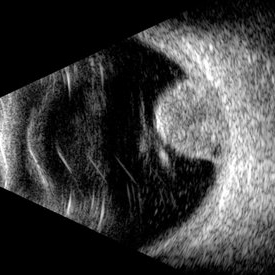
This B-mode transverse ultrasound scan reveals a mushroom-shaped choroidal tumor in the inferior nasal quadrant adjacent to the optic nerve head. The lesion appears solid with homogeneous internal reflectivity and is associated with minimal surrounding subretinal fluid and scleral excavation. It measures 6.54 mm in height × 7.52 mm in base diameter (transverse view) and extends 9.52 mm longitudinally. The vitreous contains abundant punctate opacities consistent with pigment dispersion. The retina and choroid remain attached elsewhere.
Photographer: Gustavo U. Fonseca Aguirre, Hospital Conde de Valenciana, Ciudad de México
Condition/keywords: choroidal melanoma
-
 choroidal nevus with polypoidal choroidal vasculopathy
Nov 20 2012 by Roy Schwartz, MD
Rare combination of a choroidal nevus complicated by polypoidal choroidal vasculopathy. The lesion is temporal to the fovea, and leakage of subretinal fluid almost reaching the fovea is demonstrated.
Photographer: Galit Yair-Pur
Condition/keywords: choroidal nevus, polypoidal choroidal vasculopathy (PCV)
-
 Chronic Central Serous Chorioretinopathy (CSCR)
Mar 31 2025 by Niloofar Piri, MD
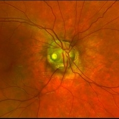
Fundus Autofluorescence image of the right eye demonstrates classic guttering with hyper autofluorescence consistent with chronic CSCR. Guttering occurs where subretinal fluid migrates inferiorly due to gravity and stressed RPE cells accumulate lipofuscin material from high turnover of photoreceptor outer segments.
Photographer: Stefan Raev, COT; Saint Louis University
Condition/keywords: central serous chorioretinopathy (CSCR), Chronic CSR, Guttering
-
 CNV due to AMPPE
Oct 16 2012 by Ratimir Lazic, MD, PhD
Color fundus photography of a 58-year-old male. White dots with juxtafoveolar subretinal fluid can be seen. BCVA of that eye is 0.35.
Photographer: Marko Lukic, MD
Imaging device: Zeis Visucam Lite 2
Condition/keywords: acute posterior multifocal placoid pigment epitheliopathy (APMPPE), choroidal neovascularization (CNV)
-
 Colobomatous Optic Disc Maculopathy
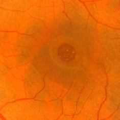
Feb 13 2020 by Yoshihiro Yonekawa, MD, FASRS
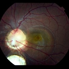
Beautifully focused fundus photograph of a teenage girl with submacular fluid from a colobomatous optic disc.
Photographer: Netanya Lerner, COA, Wills Eye Hospital/Mid Atlantic Retina
Imaging device: Topcon
Condition/keywords: chorioretinal coloboma, coloboma of optic disc, congenital optic nerve pit, subretinal fluid
-
 Colobomatous Optic Disc Maculopathy
Feb 13 2020 by Yoshihiro Yonekawa, MD, FASRS
Fluorescein angiography, late frame, of a teenage girl with submacular fluid from a colobomatous optic disc. The camera is focused is on the elevated macula, and the disc is subtly defocused.
Photographer: Netanya Lerner, COA, Wills Eye Hospital/Mid Atlantic Retina
Imaging device: Topcon
Condition/keywords: chorioretinal coloboma, coloboma of optic disc, congenital optic nerve pit, subretinal fluid
-
 Dome-Shaped Macula With Subretinal Fluid
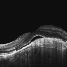
Jun 14 2018 by Gerardo Garcia-Aguirre, MD
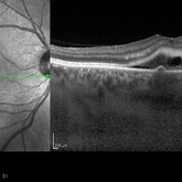
EDI OCT of the right eye of a 17-year-old highly myopic girl. Subfoveal fluid is present. There is choroidal thinning, and scleral thickening in the foveal area.
Photographer: Gerardo Garcia-Aguirre, MD
Imaging device: Heidelberg Spectralis
Condition/keywords: dome shaped macula, high myopia
-
 Hemangioma of Retina
Mar 5 2025 by Virginia Gebhart
64 year old male with choroidal hemangioma in the macula and STA. Persistent IRF and new cuff of SRF compared to previous photos. BCVA CF@face. Pt has had PDT in the past with no significant improvement. Will observe closely
Photographer: Virginia Gebhart, Retina Consultants of Carolina
Imaging device: Optos California
Condition/keywords: hemangioma, inferior subretinal fluid
-
 Hemangioma of Retina (FAF)
Mar 5 2025 by Virginia Gebhart
Fundus autofluorescence of 64 year old male with choroidal hemangioma in the macula and STA. Persistent IRF and new cuff of SRF compared to previous photos. BCVA CF@face. Pt has had PDT in the past with no significant improvement. Will observe closely
Photographer: Virginia Gebhart, Retina Consultants of Carolina
Imaging device: Optos California
Condition/keywords: autofluorescence imaging, hemangioma, inferior subretinal fluid
-
 HRA -OCT of left eye Choroidal Osteoma
Jun 2 2018 by awaneesh m upadhyay, MBBS, DNB
23-year-old male patient's left eye OCT showing undulating surface of osteoma with subretinal fluid and shaggy RPE.
Photographer: Hiteshwar Saikia
Imaging device: Heilderberg spectralis
Condition/keywords: choroidal osteoma
-
 Idiopathic Uveal Effusion Syndrome
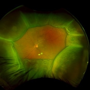
Jun 13 2023 by Ahmad B. Tarabishy, MD
66 year old male presented with a 4 month vision of painless decreased vision in the left eye. Clinical findings consistent with idiopathic uveal effusion syndrome. Fundus photography shows 360 degree choroidal elevation with dependent inferior subretinal fluid.
Photographer: Dr. Angela Rico, Retina Specialists of Tampa
Imaging device: Idiopathic Uveal Effusion Syndrome
Condition/keywords: idiopathic uveal effusion syndrome, uveal effusion
-
 Macular Hole
Mar 29 2013 by Henry J. Kaplan, MD
Chronic macular hole with drusen like deposits and surrounding cuffing of subretinal fluid.
Condition/keywords: macular hole
-
Macular Hole Repair Under PFCL
Jun 6 2024 by DAVID PÉREZ GONZÁLEZ, MD
The heavy liquid, though slightly more expensive, can be a valuable tool for macular hole repair. When applied to the posterior area, it can stabilize the hole's edges, facilitating its position and draining the adjacent subretinal fluid.
Condition/keywords: ILM peeling, macular hole, Perfluorocarbon Liquid, PFCL, vitrectomy
-
 Optic Nerve Pit Right Eye
Feb 15 2021 by Kim Barrett
A 14-year-old male presented with vision loss and VF defect. Patient was treated for presumed amblyopia with patching since age 4. He has had neurologic care for post traumatic skull fracture and brain bleed in 2012. IOP's WNL. OD is without retinoschisis or subretinal fluid. Patient is at risk of serous detachment. Current VA OD 20/200+1 PH 20/80.
Photographer: Kim Barrett C.O.A. Retina Specialist of Michigan, Grand Rapids, MI
Imaging device: Optos California
Condition/keywords: amblyopia, hemifield, Humphrey visual field, nerve, optic nerve pit, visual field defect
-
 Recurrent Central Serous Choroidopathy
Aug 21 2012 by Edwin H. Ryan, MD
EDI-OCT showing thickened choroid and subretinal fluid
Photographer: Edwin Ryan Jr. MD, VitreoRetinal Surgery, PA
Imaging device: Heidelberg Spectralis
Condition/keywords: central serous chorioretinopathy (CSCR), choroidal thickening, enhanced depth imaging
-
 Vitreomacular Traction
Jun 15 2022 by Zach Seim
Optical Coherence Tomography (OCT) of a 69 year old male with Vitreomacular Traction affecting his right eye. Patient was referred to this office for Choroidal Melanoma in his right eye in May 2021. The patient was treated with Brachytherapy in July 2021 and this OCT was taken at a follow-up appointment in May 2022. Patient's vision was 20/30-2 at the time this OCT was taken. Patient states that his vision was better since his last visit, and that he sees floaters occasionally.
Photographer: Zach Seim
Imaging device: Heidelberg Spectralis
Condition/keywords: heidelberg spectralis, OD, optical coherence tomography (OCT), right eye, subretinal fluid, vitreomacular adhesion, vitreomacular interface disorders, vitreomacular traction (VMT)
 Loading…