Search results (91 results)
-
 The Peripheral Retina in Profile: A Stereoscopic Atlas
Mar 12 2013 by Norman Byer
The stereoscopic atlas contains unique stereo photographs vividly portraying the changes in the peripheral fundus and their histopathology, incidence and risks.
Condition/keywords: stereo pair, video
-
 Inferior Choroidal Coloboma and Tilted Disc
Feb 19 2013 by From the Collections of Thomas M. Aaberg, MD and Thomas M. Aaberg Jr., MD
NLP; Left of stereo pair.
Condition/keywords: coloboma, stereo pair
-
 Lattice Degeneration
Jan 5 2015 by H. Michael Lambert, MD
Lattice degeneration (stereo pair B).
Condition/keywords: lattice degeneration
-
 Lattice Degeneration
Jan 5 2015 by H. Michael Lambert, MD
Retinal detachment with lattice degeneration and holes (stereo pair A).
Condition/keywords: lattice degeneration
-
 Coloboma, In Stereo
Oct 1 2012 by Michael P. Kelly, FOPS
This is a stereo retinal fundus photograph of a coloboma, with the optic nerve centered, using a Zeiss FF3C retinal fundus camera.
Photographer: Michael P. Kelly, FOPS Director, Duke Eye Labs, Duke University Hospital, Duke Eye Center, Durham, NC
Condition/keywords: coloboma, fundus photograph, stereo pair
-
 Retinal Detachment, in Stereo
Oct 4 2012 by Michael P. Kelly, FOPS
Retinal detachment showing bare RPE, in stereo.
Photographer: Michael P. Kelly, FOPS, Director, Duke Eye Center Labs, Duke University Hospital, Durham, NC
Condition/keywords: stereo pair
-
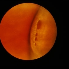
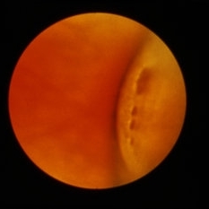
 ON pits OD stereo L
Sep 18 2012 by Pauline T Merrill, MD, FASRS
2 pits in right optic nerve of a 77-year-old woman with bilateral optic nerve pits and glaucoma, stable over 20 years (L image of stereo pair)
Photographer: Karen Parque, Illinois Retina Associates, Chicago, IL
-
 Choroidal Detachment, In Stereo
Sep 25 2012 by Michael P. Kelly, FOPS
Photographer: Michael P. Kelly, FOPS Director, Duke Eye Labs, Duke University Hospital, Duke Eye Center, Durham, NC
Imaging device: Zeiss FF3C
Condition/keywords: choroidal detachment, stereo pair
-
 Inferior Choroidal Coloboma and Tilted Disc
Feb 19 2013 by From the Collections of Thomas M. Aaberg, MD and Thomas M. Aaberg Jr., MD
NLP; Right of stereo pair.
Condition/keywords: coloboma, stereo pair
-
 ON pit OS stereo L
Sep 18 2012 by Pauline T Merrill, MD, FASRS
Left optic nerve of a 77-year-old woman with bilateral optic nerve pits and glaucoma, stable over 20 years (L image of stereo pair)
Photographer: Karen Parque, Illinois Retina Associates, Chicago, IL
-
 Proliferative Diabetic Retinopathy, In Stereo
Sep 28 2012 by Michael P. Kelly, FOPS
Proliferative diabetic retinopathy, PDR, Stereo.
Photographer: Michael P. Kelly, FOPS Director, Duke Eye Labs, Duke University Hospital, Duke Eye Center, Durham, NC
Imaging device: Zeiss FF4
Condition/keywords: stereo pair
-
 Asteroid Hyalosis, In Stereo
Sep 28 2012 by Michael P. Kelly, FOPS
Asteriod hyalosis, stereo.
Photographer: Michael P. Kelly, FOPS Director, Duke Eye Labs Duke University Hospital, Duke Eye Center, Durham, NC
Imaging device: Zeiss FF3C
Condition/keywords: asteroid hyalosis, stereo pair
-
---thumb.jpg/image-square;max$300,300.ImageHandler) Chronic Central Serous Chorioretinopathy
Chronic Central Serous Chorioretinopathy
Feb 20 2013 by From the Collections of Thomas M. Aaberg, MD and Thomas M. Aaberg Jr., MD
Stereo pair color fundus photo of OD of probable bilateral chronic CSR at the macula.
Condition/keywords: chronic central serous chorioretinopathy (CSCR), color photo, stereo pair
-
---thumb.jpg/image-square;max$300,300.ImageHandler) Chronic Central Serous Chorioretinopathy
Chronic Central Serous Chorioretinopathy
Feb 20 2013 by From the Collections of Thomas M. Aaberg, MD and Thomas M. Aaberg Jr., MD
Stereo pair color fundus photo of OS of probable bilateral chronic CSR at the macula.
Condition/keywords: color photo, stereo pair
-
---thumb.jpg/image-square;max$300,300.ImageHandler) Chronic Central Serous Chorioretinopathy
Chronic Central Serous Chorioretinopathy
Feb 20 2013 by From the Collections of Thomas M. Aaberg, MD and Thomas M. Aaberg Jr., MD
Stereo pair color fundus photo of OS of probable bilateral chronic CSR at the macula.
Condition/keywords: chronic central serous chorioretinopathy (CSCR), color photo, stereo pair
-
 Diabetes Proliferative
Jul 11 2013 by Jerald A. Bovino, MD
No history, traction retinal detachment, part of stereo pair.
Condition/keywords: diabetic traction detachment
-
 Diabetes Proliferative
Jul 11 2013 by Jerald A. Bovino, MD
No history, traction retinal detachment, part of stereo pair.
Condition/keywords: diabetic mellitus, stereo pair, tractional retinal detachment
-
 Diabetic Retinopathy Optic Nerve Edema, Fluorescein Angiogram, Stereo
Apr 11 2015 by James B. Soque, CRA, OCT-C, COA, FOPS
Optic Nerve Edema and Leakage on fluorescein angiography in this 48-year-old patient with a 10 year history of diabetes. 50 degree stereo photo fluorescein angiogram.
Photographer: James B. Soque, CRA, COA
Imaging device: Topcon TRC 50 DX, OIS 5 MP Digital Camera, MERGE Software
Condition/keywords: background diabetic retinopathy (BDR), diabetes, disc leakage, fluorescein leakage, optic disc swelling, optic nerve edema, stereo pair
-
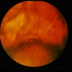
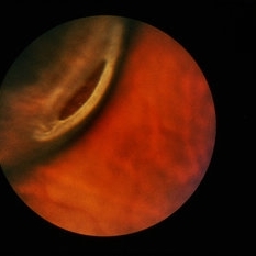
 Horseshoe Tear, In Stereo
Sep 28 2012 by Michael P. Kelly, FOPS
Horse shoe tear, stereo.
Photographer: Michael P. Kelly, FOPS Director, Duke Eye Labs, Duke University Hospital, Duke Eye Center, Durham, NC
Imaging device: Zeiss FF3C
Condition/keywords: stereo pair
-
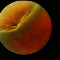
 Lattice Degeneration
Jan 5 2015 by H. Michael Lambert, MD
Lattice degeneration with holes and small retinal detachment (stereo pair A).
Condition/keywords: lattice degeneration
-
 Lattice Degeneration
Jan 5 2015 by H. Michael Lambert, MD
Lattice degeneration with holes and small retinal detachment (stereo pair B).
Condition/keywords: lattice degeneration
-
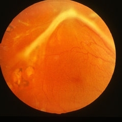
 Lattice Degeneration
Jan 5 2015 by H. Michael Lambert, MD
Lattice degeneration with large atrophic hole (stereo pair A).
Condition/keywords: lattice degeneration
 Loading…