Search results (28 results)
-
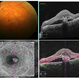
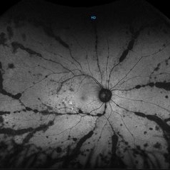 Autofluorescence Stage 3 Vogt-Koyanagi-Harada (VKH) Disease
Oct 20 2021 by Bryon R McKay, MD, PhD, FRCSC, DRCPSC - Retina
27yF presented with sub-acute findings of VKH, she has an interesting pattern of perivascular changes. She was successfully treated with immunosuppressive agents and maintains 20/20 vision.
Photographer: Dr. K. Vaezi, University of British Columbia, Canada
Imaging device: Optos Imaging system
Condition/keywords: Vogt-Koyanagi-Harada
-
 Myopic Traction Maculopathy
Mar 17 2025 by Drew Mitchell
HD 1 line 100x 9 mm scan of a right eye with MTM at stage 3c. Macular Schisis Detachment.
Photographer: Drew Mitchell OCT-C
Imaging device: Zeiss Cirrus 5000
Condition/keywords: full thickness macular hole, Macular hole, myopic foveoschisis, myopic macular schisis, myopic traction maculopathy, PVD
-
 Coats' Disease - Stage 3A
Aug 21 2019 by Victor M Villegas, MD
Coats' Disease - stage 3A.
Condition/keywords: abnormal retina, Coats' disease, diffuse lipid exudate, edema, foveal hard exudates, pediatic retina, retcam, retinal angioma
-
 Retinopathy of Prematurity
Jul 12 2021 by Stefanie Palmer
Retinopathy of prematurity Stage 3 in a 5 month old baby. The flying baby technique was used to create this image.
Photographer: Stefanie Palmer CRA
Imaging device: scanning laser ophthalmoscope
Condition/keywords: retinopathy of prematurity (ROP), retinopathy of prematurity stage 3
-
 Sickle Cell Retinopathy
Feb 15 2021 by Kim Barrett
24-year-old female with Sickle Cell Retinopathy, stage 3. She confirms she has the trait as well as her grandmother, mother and a sibling. She has seafan neovascularization superotemporal OD. Current VA is 20/20. Photo is pre-PRP laser with areas of non-profusion temporally.
Photographer: Kim Barrett C.O.A. Retina Specialist of Michigan, Grand Rapids, MI
Imaging device: Optos California
Condition/keywords: neovascularization (NV), pan-retinal photocoagulation (PRP), sickle cell retinopathy, stage 3, trait
-
 Proliferative Sickle Retinopathy Stage 3
Oct 9 2012 by Alan D. Letson, MD
21-year-old man with Hgb SC Disease and stage 3 PSR with autoinfarction of sea fans
Photographer: Beverly Radcliffe
Condition/keywords: autoinfarction, sea fan, sickle cell, sickle cell retinopathy
-
 ROP
ROP
Mar 26 2025 by Korey Starkey
9 month old patient presents today with Retinopathy of Prematurity in both eyes. Patient was born at gestational age of 25 weeks 2 days, 940g. Left eye presents with vitreoretinal traction and peripheral VH with regressed stage 3 and persistent stage 2 disease.
Photographer: Korey Starkey
Imaging device: Optos
Condition/keywords: retinopathy of prematurity stage 2, rop, stage 3, vitreoretinal traction, vitreous hemorrhage
-
 Stage 3 ROP
Aug 20 2018 by Anna L. Ells, MD, FRCS(C)
Posterior zone II, stage 3 ROP with plus disease
Photographer: Anna Ells, Calgary Retina Consultants
Imaging device: RetCam
Condition/keywords: retinopathy of prematurity stage 3
-
 High Magnification of Stage 3 Neovascularization in ROP
Aug 18 2018 by Anna L. Ells, MD, FRCS(C)
High magnification of stage 3 neovascularization in ROP.
Photographer: Leslie MacKeen
Imaging device: RetCam
Condition/keywords: retinopathy of prematurity stage 3
-
 Stage 3 Vogt-Koyanagi-Harada (VKH) Disease
Oct 20 2021 by Bryon R McKay, MD, PhD, FRCSC, DRCPSC - Retina
27yF presented with sub-acute findings of VKH, she has an interesting pattern of perivascular changes. She was successfully treated with immunosuppressive agents and maintains 20/20 vision.
Photographer: Dr. Vaezi, University of British Columbia
Imaging device: Optos Imaging System
Condition/keywords: uveitis
-
 High Magnification of Stage 3 Neovascularization
Aug 18 2018 by Anna L. Ells, MD, FRCS(C)
High magnification highlighting neovascularization of stage 3 ROP. Note the "popcorn" just posterior to the neovascularization.
Photographer: Leslie Mackeen
Imaging device: Retcam
Condition/keywords: retinopathy of prematurity stage 3
-
 Stage 3 ROP
Aug 18 2018 by Anna L. Ells, MD, FRCS(C)
Left retinal image of premature infant born at 24 weeks gestational age, birthweight of 653 grams. Image taken at 38 weeks of post-menstrual age. Zone II, stage 3 ROP. Moderate plus disease. Good example of Type I ROP requiring treatment.
Photographer: Leslie Mackeen, Hospital for Sick Children, Toronto, Canada
Imaging device: RetCam
Condition/keywords: retinopathy of prematurity stage 3
-
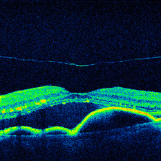 OCT Cirrus HD 5 Line Scan ARMD SRF RPED Stage 3 PVD
Mar 6 2013 by James B. Soque, CRA, OCT-C, COA, FOPS
Zeiss Cirrus OCT 4000, Hi Definition 5 Line Scan, 91-year-old white female with peripapillary SRN with subretinal heme, serous fluid, and a stage 3 PVD, still attached at the optic nerve.
Photographer: James Soque, CRA, COA, Island-Retina
Imaging device: Zeiss Cirrus 4000 SD OCT with 6.0.2.81 Software
Condition/keywords: optical coherence tomography (OCT)
-
 Retinopathy of Prematurity Stage 3
Oct 23 2012 by Larry Halperin, MD
Retinopathy of prematurity, stage 3
Condition/keywords: retinopathy of prematurity (ROP), retinopathy of prematurity stage 3
-
 Stage 3 Macular Hole
-
 Macular Hole
Sep 20 2012 by Jeffrey G. Gross, MD, FASRS
Macular hole, Stage 3, pre-op 20/400
Condition/keywords: macular hole, pre-op
-
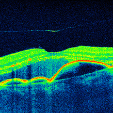 OCT Cirrus 5 Line HD Scan EDI ARMD SRF RPED Stage 3 PVD
Mar 6 2013 by James B. Soque, CRA, OCT-C, COA, FOPS
Zeiss Cirrus OCT 4000, EDI Aquired Using Enhanced Depth Mode, 91-year-old white female with peripapillary SRN with subretinal heme, serous fluid, and a stage 3 PVD, still attached at the optic nerve.
Photographer: James Soque, CRA, COA, Island-Retina
Imaging device: Zeiss Cirrus 4000 SD OCT with 6.0.2.81 Software
Condition/keywords: optical coherence tomography (OCT)
-
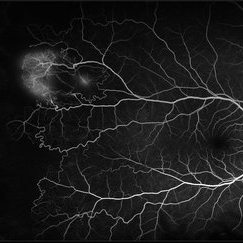
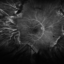 Fluorescein Angiogram of ROP With Cryo Scarring
Jul 7 2025 by Jenn Geelan
FA photo of a 34 year old male with prior stage 3 ROP with history of 360 degree cryotherapy.
Photographer: Jenn Geelan, Retina-Vitreous Surgeons of CNY
Imaging device: Optos California
Condition/keywords: cryotheraphy scar, fluorescein angiogram (FA), fundus photograph, retinopathy of prematurity (ROP), ROP, tilted disc
-
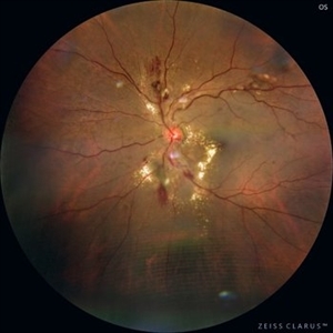 Hypertensive Retinopathy
Sep 16 2025 by Píndaro Alonso Cruz-Benitez
Hypertensive Retinopathy Stage 3 Male 55 year-old
Photographer: Pindaro Alonso Cruz Benitez
Condition/keywords: exudate, Haemorrhage, hypertensive retinopathy
-
 Macular Hole Stage 3
Sep 27 2012 by Jeffrey G. Gross, MD, FASRS
Macular hole stage 3 post op with gas bubble 20/60.
Condition/keywords: 20/60, gas bubble, macular hole, post-op
-
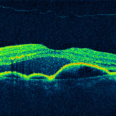 OCT Cirrus 5 Line Scan ARMD SRF RPED Stage 3 PVD
Mar 6 2013 by James B. Soque, CRA, OCT-C, COA, FOPS
Zeiss Cirrus OCT 4000, 5 Line Scan , 91-year-old white female with peripapillary SRN with subretinal heme, serous fluid, and a stage 3 PVD, still attached at the optic nerve.
Photographer: James Soque, CRA, COA, Island-Retina
Imaging device: Zeiss Cirrus 4000 SD OCT with 6.0.2.81 Software
Condition/keywords: optical coherence tomography (OCT)
-
 Retinal Angiomatous Proliferation
May 7 2021 by Dhaivat Shah
Retinal angiomatous proliferation (RAP) is a distinct variant of neovascular age-related degeneration (AMD) that usually initiates at the retina and progresses posteriorly into sub retinal space. In most recent study, it was suggested that angiogenesis may begin in the retina, choroid, or both, and introduced a new name for the process: Type 3 neovascularization. The frequency of RAP has been studied in many studies, with figures ranging from 10% to 21% of exudative AMD. Clinically, three stages were originally described as intraretinal neovascularization (IRN), subretinal neovascularization (SRN), choroidal neovascularization (CNV). RAP predominantly intraretinal hard exudates, and intra/pre retinal hemorrhages along with intraretinal edema, associated pigment epithelial detachment beneath it, at times retinochoroidal, retino-retinal anastomosis. Apart from conventional OCT, FFA and ICG, OCT-A has now been used primarily as a tool in the diagnosis RAP. Here we present imaging of a 30-year-old young male diagnosed as RAP stage 3 (Type 3 CNVM). Patient was started on intravitreal anti-VEGF monotherapy therapy.
Photographer: Choithram Netralaya Indore
Condition/keywords: retinal angiomatous proliferation (RAP)
-
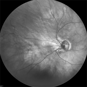 ROP stage 3
Sep 22 2022 by Filip Kecer
IR cSLO widefield image of an 27-year-old man born as premature
Photographer: Filip Kecer, National Institute of Childrens Diseases
Imaging device: Spectralis, Heidelberg Engineering
Condition/keywords: retinopathy of prematurity stage 3, rop
-
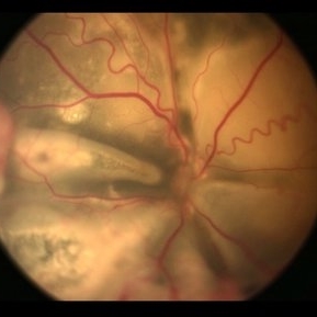
 ROP-Zone-I-Stage-3-Plus
Jun 3 2022 by Dipak Nag, MBBS, FCPS, MSc, FRF
Fundus photograph of a child of gestational age 26 weeks and birth weight 1050 grams, shows dilatation and tortuosity of vessels in zone I, extra-retinal fibro-vascular proliferation, hemorrhage with huge peripheral avascular area.
Photographer: Dipak Nag, National Institute of Ophthalmology, Dhaka, Bangladesh
Imaging device: RetCam shuttle
Condition/keywords: retinopathy of prematurity (ROP), retinopathy of prematurity Plus disease, retinopathy of prematurity stage 3, retinopathy of prematurity zone I
-
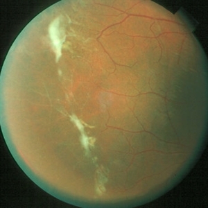
 Stage 3 Coats' Disease
Aug 7 2022 by Muhammad Amer Awan, MD, FRCSEd, FRCOphth, FRCS Glasgow, FACS, FASRS
Fundus photography of a 6 months old baby boy who presented with unilateral leucoria. There was right exudate retinal detachment with extensive hard exudates and tortuous retinal vessels. Diagnosis of Coats' disease was made that was externally drained and intravitreal rhanibizumab was given.
Photographer: Muhammad Amer Awan, Shifa Taamer e Millat University
Condition/keywords: Coats' disease, exudative retinal detachment, exudative retinopathy, unilateral exudative retinal detachment
 Loading…