Search results (6 results)
-
 ERM with Retinal Striae
Apr 13 2023 by Virginia Gebhart
Right eye fundus photo of 65-year-old male with severe ERM with retinal striae s/p bilateral RD repair
Photographer: Virginia Gebhart, Retina Consultants of Carolina
Imaging device: Topcon TRC 50DX
Condition/keywords: epiretinal membrane (ERM), retinal strial
-
 Multi-Color Image of Epiretinal Membrane With Prominent Edge Superior Temporal Macula
Nov 7 2018 by Tammy Mclaughlin
Multi-color image of female patient with epiretinal membrane OD. Clinical exam and diagnostic testing show large central ERM, retinal striae, and a prominent edge in the superior temporal macula. Visual acuity 20/25.
Photographer: Tammy Mclaughlin, Carolina Retina Center, Sumter SC 29150
Imaging device: Heidelberg
Condition/keywords: epiretinal membrane (ERM)
-
 Partial Optic Disc Avulsion with Optic Disc Pit
Jul 1 2018 by John S. King, MD
16-year-old with acute loss of vision after blunt finger injury to eye while playing football. This photo is three weeks post-injury. Vision HM. Retinal striae with subhyaloid heme. Decreased retinal whitening. Peripapillary heme clearing, and temporal optic disc avulsion with optic disc pit can be seen.
Photographer: Maisee Yang
Imaging device: Topcon
Condition/keywords: epiretinal membrane (ERM), optic nerve head avulsion, optic nerve pit, traumatic optic neuropathy
-
 Pinching a Stained ILM
Feb 4 2022 by Manish Nagpal, MD, FRCS (UK), FASRS
ILM peeling initiated by carefully pinching the surface of retina around the macular hole revealing radiating striae confirming the right plane.
Photographer: Manish Nagpal, Director, Retina Foundation, Ahmedabad
Imaging device: Sony PMW -10 MD surgical camera
Condition/keywords: ILM flap, ILM staining, internal limiting membrane (ILM) peeling, macular hole, retinal striae
-
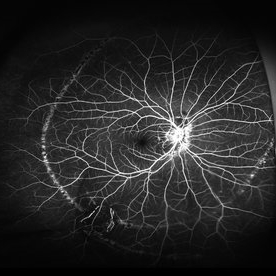 Schlaegel Line
Mar 14 2024 by César Adrián Gómez Valdivia, MD
Hyperfluorescent, concentric, chorioretinal striae on fluoroangiography.
Photographer: Erika Paulina Ornelas Cazares
Imaging device: California ICG OPTOS
Condition/keywords: line, PHOS, Schlaegel
-
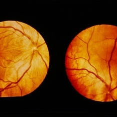 Vogt-Koyanagi-Harada Disease
Feb 20 2015 by H. Michael Lambert, MD
No history. Color photo showing multiple retinal detachments throughout the posterior right eye with prominent retinal striae. There is possible swelling of the optic nerve. The optic nerve may be swollen.
Condition/keywords: exudative retinal detachment, Vogt-Koyanagi-Harada
 Loading…