Search results (10 results)
-
 PEHCR (Peripheral Exudative Hemorrhagic Chorioretinopathy)
May 12 2023 by Niloofar Piri, MD
Ultrawide fundus photograph of the left eye demonstrating extensive peripheral hemorrhagic exudative detachment in a 79 yo Caucasian female with prior history of non-exudative AMD. Recent diagnosis of Acute myeloid leukemia with low platelet count which might have contributed to the above presentatuon. Please note the temporal subretinal hemorrhage as well as RPE atrophy and hyperplasia in the macula.
Photographer: Rocio Bentivegna, MD, Saint Louis University; Jessica Maddox, COA, Saint Louis University
Condition/keywords: peripheral exudative hemorrhagic chorioretinopathy (PEHCR)
-
 Classic presentation of PEHCR in an elderly Asian female
Apr 15 2024 by David A Reichstein, MD
(A) Ultra-widefield color fundus photograph demonstrating a large, localized area of subretinal fluid surrounded by lipid exudation at its superior and posterior borders. (B) Posterior segment B-scan ultrasonography demonstrates that the lesion is hollow, suggestive of localized subretinal fluid. (C) Early-stage ultra-widefield FA demonstrates an absence of early fluorescence. (D) Late-stage ultra-widefield FA demonstrates late hyperfluorescence.
Condition/keywords: peripheral exudative hemorrhagic chorioretinopathy (PEHCR)
-
 Large PEHCR causing an exudative inferior detachment in a patient with AMD
Apr 15 2024 by David A Reichstein, MD
(A) Ultra-widefield color fundus photograph demonstrates a temporal PEHCR causing minimal intra- and subretinal hemorrhage along with lipid exudation. There is an associated inferior detachment due to the dependent nature of the exudation. Note the lipid exudation at the posterior edge of the detachment indicating chronicity of the lesion. Drusen in the macula are also appreciated. (B) Ultra-widefield FA in early stage demonstrates hypofluorescence temporally and inferiorly. (C) Ultra-widefield color fundus photograph taken after 1 year of monthly anti-VEGF therapy demonstrates resolution of the exudative detachment and resultant chorioretinal scarring.
Condition/keywords: peripheral exudative hemorrhagic chorioretinopathy (PEHCR)
-
 Macula-threatening PEHCR causing a nasal visual-field defect in a patient with AMD
Apr 15 2024 by David A Reichstein, MD
(A) Ultra-widefield color fundus photograph demonstrates a PEHCR encroaching upon the temporal macula. Lipid exudation is apparent at the lesion’s anterior and inferior border. Subretinal hemorrhage is apparent at the lesion’s inferior border. Drusen are apparent in the macula. An unrelated, small choroidal nevus is apparent in the inferior fundus. (B) Ultra-widefield FA taken in early stage demonstrates hypofluorescence within the lesion consistent with blockage by possible sub-RPE or subretinal heme. (C) Ultra-widefield fundus photograph taken 6 months following the initiation of monthly anti-VEGF therapy demonstrates considerable reduction in the size of the lesion and resolution of the subretinal hemorrhage and lipid exudation. (D) Ultra-widefield fundus photograph taken 1 year after presentation where a treat-and-extend approach was performed for the most recent 6 months. The lesion had almost completely resolved.
Condition/keywords: peripheral exudative hemorrhagic chorioretinopathy (PEHCR)
-
 PEHCR
PEHCR
Jan 4 2024 by Virginia Gebhart
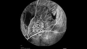
86 year old male with partially oxidized choroidal hemorrhage and CME. Previous FA shows blocking defect temporally, most likely a choroidal hemorrhage with SRH and late leakage. Continued improvement with 8 week intervals of Eylea. VA 20/60 Previous RD repair with scleral buckle and cryo in 1980's
Photographer: Virginia Gebhart
Imaging device: Optos California
Condition/keywords: chorioretinopathy, choroidal hemorrhage, cystoid macular edema (CME), peripheral exudative hemorrhagic chorioretinopathy (PEHCR)
-
 Peripheral Exudative Hemorrhagic Chorioretinopathy
Oct 7 2020 by Olivia Rainey
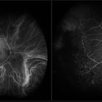
Fluorecein and ICG angiography of a 80-year-old male with peripheral exudative hemorrhagic chorioretinopathy affecting his right eye. Patient noted only mild floaters for a couple weeks OD on 10/5/2020. The physician strongly suspects that the lesion is subretinal blood (likely from PEHCR) rather than choroidal melanoma. There is blocking on FA and ICG with a definite lack of intrinsic vessels within the mass lesion. He will monitor closely, as the patient is monocular with a history of multiple surgeries (which the family believes PPV for "scar tissue") OS mostly in 2013. His family also reports remembering possibly being told there was a "small mass" in the left eye at one point in their surgical course. The physician believes that it's possible this was a bleed related to PEHCR as it typically exists as a bilateral condition.
Photographer: Olivia Rainey, OCT-C, COA
Imaging device: Optos California
Condition/keywords: fluorescein angiogram (FA), indocyanine green (ICG) angiography, monocular, Optos, peripheral exudative hemorrhagic chorioretinopathy (PEHCR), periphery, subretinal hemorrhage, ultra-wide field imaging
-
 Peripheral Exudative Hemorrhagic Chorioretinopathy
Mar 9 2023 by Christopher R. Adam, M.D.
72 year-old Caucasian woman referred for possible ocular melanoma. Exam and multimodal imaging including ultrasound and angiography showed bilateral hemorrhagic peripheral lesions an pigmentary degeneration consistent with PEHCR.
Condition/keywords: peripheral exudative hemorrhagic chorioretinopathy (PEHCR)
-
 Peripheral Exudative Hemorrhagic Chorioretinopathy
Nov 19 2024 by Toolie Winters
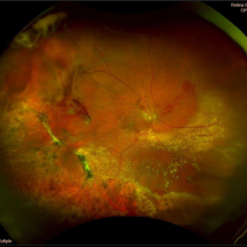
Ultra-wide field fundus photograph of an 85-year-old woman with Peripheral Exudative Hemorrhagic Chorioretinopathy (PECHR) affecting the right eye. Patient presented with a blind spot centrally in the right eye which she first noticed 4 months prior to this image being taken. The patient states that in the month prior to this image, she began noticing bright lights flash across her vision 4-5x/day which last about 15 seconds. The flashes are either black with a blue ring around them or yellow, and their frequency has increased over time. The patient's vision at the time of this appointment was Dcc20/100+1 PHNI. This photo also shows diffuse hemorrhage, lipid, and an eccentric disciform lesion.
Photographer: Toolie Winters
Imaging device: Optos California
Condition/keywords: fundus photography, neovascular age-related macular degeneration (AMD), Optos, OPTOS CALIFORNIA, peripheral exudative hemorrhagic chorioretinopathy (PEHCR), pseudocolor, ultra-wide field imaging, wet age-related macular degeneration (wet AMD)
-
 Peripheral Polypoidal Choroidal Vasculopathy Causing PEHCR
Aug 25 2020 by Kshitij Raizada, MS Ophthalmology
A 36-year-old female presented to with complaints of diminution of vision in LE for 3 months. Her BCVA in the RE was 6/6 and CF@1m in the LE. She was a K/C/O polypoidal choroidal vasculopathy (PCV) in the RE and had a history of receiving 2 doses of intravitreal Aflibercept (Eylea) in the RE. On her visit, she had dense Vitreous hemorrhage in the LE. 25G pars plana vitrectomy + intravitreal Aflibercept was planned for her. On clearing the vitreous hemorrhage, the patient was found to have Peripheral Exudative Hemorrhagic Chorioretinopathy (PEHCR). An on-table diagnosis of "PCV causing PEHCR" was made. Endolaser was done to sites suspicious to have underlying polyps. The patient's vision improved to 6/18 in the LE after one week of surgery. One month post-surgery, her BCVA in the LE had improved to 6/9. ICG angiography was done which revealed non-leaking BVN(branching vascular network) and no polyps. The patient has been doing well and has been kept under observation.
Condition/keywords: indocyanine green (ICG) angiography, peripheral exudative hemorrhagic chorioretinopathy (PEHCR), polypoidal choroidal vasculopathy (PCV), video
-
Pseudomelanoma (PEHCR)
Apr 15 2025 by Virginia Gebhart
67 year old male referred for peripheral choroidal lesion. Clinical exam and Bscan findings consistent with a subRPE hemorrhage secondary to peripheral exudative hemorrhagic chorioretinopathy. No vascularity on ultrasound. OD has small subRPE hemorrhage as well. Pt is on Eliquis. Will monitor with serial exams. Sponsored by the number 2
Photographer: Virginia Gebhart, Retina Consultants of Carolina
Imaging device: Optos California
Condition/keywords: peripheral exudative hemorrhagic chorioretinopathy (PEHCR)
 Loading…