Search results (19 results)
-
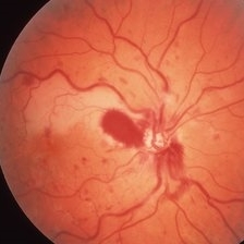
 Commotio Retinae with Retinal Hemorrhages
Mar 27 2018 by Nichole Lewis
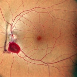
14-year-old male hit in the right eye with a stick. Commotio Retinae with retinal hemorrhages and peripapillary hemorrhage.
Photographer: Nichole Lewis
Condition/keywords: commotio retinae, peripapillary hemorrhage, retinal hemorrhage
-
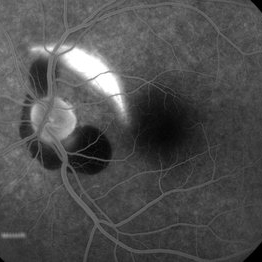 Choroidal rupture and peripapillary hemorrhage - FA
Jan 26 2013 by Roy Schwartz, MD
A 36-year-old male presented to the ER after blunt trauma to his left eye. On FA a chroidal rupture (hyperfluorescent area) was seen as well as peripapillary hemorrhage (hypofluorescent).
Photographer: Galit Yair-Pur
Condition/keywords: choroidal rupture, peripapillary hemorrhage
-
 Papilledema
Sep 21 2012 by Suber S. Huang, MD, MBA, FASRS
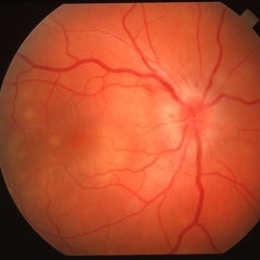
Fundus photograph of a 24-year-old obese woman with severe papilledema secondary to idiopathic intracranial hypertension.
Condition/keywords: dilated tortuous vessels, exudate, idiopathic intracranial hypertension, Paton's lines, peripapillary hemorrhage, pseudotumor cerebri
-
 10 Days Post Subretinal TPA
Jan 9 2019 by John S. King, MD
76-year-old white male with history of treat/extend with Eylea OD for a PPCNVM; also monocular due to large scar in fellow eye. Two months since last injection, had acute decrease in vision OD and was seen that day. Vision CF; moderate SRH involving the fovea. Discussed monotherapy with anti-VEGF vs displacement, and elected for PPV, srTPA, AFx, SF6. Total of 0.2 cc of 25 microgram/0.1 ml of srTPA administered from two different areas in the temporal macula. 10 days post-op vision is improving; 20/200 J7; displacement of heme (photo)
Photographer: Kay Dalby
Imaging device: Topcon 50
Condition/keywords: choroidal neovascular membrane (CNVM), peripapillary hemorrhage, presumed ocular histoplasmosis syndrome (POHS)
-
 Central Retinal Artery Occlusion
Mar 26 2019 by Gary R. Cook, MD, FACS
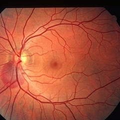
61-year-old male patient with acute CRAO OS demonstrating a hyperemic optic disc with a couple of peripapillary hemorrhages, generalized arteriolar narrowing, a cherry-red spot in the macula, and retinal whitening surrounding the fovea; VA= LP.
Imaging device: Topcon VT-50
Condition/keywords: central retinal artery occlusion (CRAO), cherry red spot, retinal whitening
-
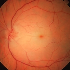
 Choroidal rupture and peripapillary hemorrhage - Fundus image
Jan 26 2013 by Roy Schwartz, MD
A 36-year-old male presented to the ER after blunt trauma to his left eye. On clinical examination peripapillary hemorrhage was seen as well as a crescent shaped area suspected for a choroidal rupture, which was confirmed in FA.
Photographer: Galit Yair-Pur
Condition/keywords: choroidal rupture, peripapillary hemorrhage
-
 Combined CRVO and BRAO
Mar 27 2019 by Gary R. Cook, MD, FACS
Right eye of a 56-year-old white male with a combined perfused CRVO (venous dilation and dot & blot hemorrhages in all 4 quadrants) and a superotemporal BRAO with peripapillary hemorrhages and cotton wool spots, and an area of retinal whitening inside of the ST arcade. V.A.= 20/70.
Imaging device: Topcon VT-50
Condition/keywords: branch retinal artery occlusion (BRAO), central retinal vein occlusion (CRVO)
-
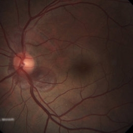
 Peripapillary Hemorrhage
Jun 10 2020 by Manish Nagpal, MD, FRCS (UK), FASRS
Peripapillary hemorrrhage.
Photographer: gayathri mohan
Imaging device: nidek slo mirante
Condition/keywords: hemorrhage
-
---thumb.JPG/image-square;max$300,300.ImageHandler) Peripapillary Hemorrhage
Peripapillary Hemorrhage
Jul 13 2013 by Jason S. Calhoun
Small optic nerve hemorrhage due to glaucoma.
Photographer: Jason S. Calhoun, Department of Ophthalmology, Mayo Clinic Jacksonville, Florida
Imaging device: TOPCON TRC 50-EX
-
 Posterior Uveitis
Apr 8 2019 by Gary R. Cook, MD, FACS
37-year-old white male with mild vitritis, optic disc hyperemia and edema, peripapillary hemorrhages and yellow-white spots in temporal macula OD; V.A. = 20/30.
Imaging device: Topcon VT-50
Condition/keywords: posterior uveitis
-
---thumb.JPG/image-square;max$300,300.ImageHandler) Radiation maculopathy
Radiation maculopathy
Nov 3 2012 by Mallika Goyal, MD
Fluorescein angiogram of an eye with radiation optic neuropathy shows hyperfluorescence of lower half of optic disc and areas of blocked fluorescence corresponding to peripapillary hemorrhages. She was treated with systemic steroids for the radiation optic neuropathy with improvement.
Photographer: Mallika Goyal
Condition/keywords: peripapillary hemorrhage, radiation optic neuropathy
-
 Subretinal Heme Involving Fovea Before PPV
Jan 9 2019 by John S. King, MD
76-year-old white male with history of treat/extend with Eylea OD for a PPCNVM; also monocular due to large scar in fellow eye. Two months since last injection, had acute decrease in vision OD and was seen that day. Vision CF; moderate SRH involving the fovea. Discussed monotherapy with anti-VEGF vs displacement, and elected for PPV, srTPA, AFx, SF6. Total of 0.2 cc of 25 microgram/0.1 ml of srTPA administered from two different areas in the temporal macula.
Photographer: Stacey Coleman
Imaging device: Topcon 50
Condition/keywords: choroidal neovascular membrane (CNVM), ocular histoplasmosis syndrome (OHS), peripapillary hemorrhage
-
 Superior Peripapillary Hemorrhage
Jul 13 2013 by Jason S. Calhoun
Patient was seen for acute vision loss in the right eye. Patient has glaucoma. VA was 20/70 in the right eye. Had vitrectomy back in May 2012 for ERM stripping. Also had trabectome with cataract surgery in December of 2012. Fundus photos presents a superior peripapillary Hemorrhage of the optic nerve. Patient will be followed up in one month.
Photographer: Jason S. Calhoun, Department of Ophthalmology, Mayo Clinic Jacksonville, Florida
Imaging device: TOPCON TRC 50-EX
Condition/keywords: peripapillary hemorrhage
-
 Superior Peripapillary Hemorrhage
Jun 27 2013 by Jason S. Calhoun
Patient was seen for acute vision loss in the right eye. Patient has glaucoma. VA was 20/70 in the right eye. Had vitrectomy back in May 2012 for ERM stripping. Also had trabectome with cataract surgery in December of 2012. Fundus photos presents a superior peripapillary Hemorrhage of the optic nerve. Patient will be followed up in one month.
Photographer: Jason S. Calhoun, Mayo Clinic Jacksonville, Florida
Imaging device: TOPCON TRC 50-EX
Condition/keywords: peripapillary hemorrhage
-
 Superotemporal Peripapillary Hemorrhage
Dec 18 2014 by H. Michael Lambert, MD
Superotemporal peripapillary hemorrhage OD following high-dive in a 15 year-old white female with peau d'orange fundus changes but biopsy negative for PXE.
Condition/keywords: superotemporal peripapillary hemorrhage
-
 Superotemporal Peripapillary Hemorrhage
Dec 18 2014 by H. Michael Lambert, MD
Superotemporal peripapillary hemorrhage OD following high-dive in a 15-year-old white female with peau d'orange fundus changes but biopsy negative for PXE.
Condition/keywords: superotemporal peripapillary hemorrhage
-
 Superotemporal Peripapillary Hemorrhage
Dec 18 2014 by H. Michael Lambert, MD
Nearly resolved superotemporal peripapillary hemorrhage OD following high-dive in a 15-year-old white female with peau d'orange fundus changes but biopsy negative for PXE.
Condition/keywords: superotemporal peripapillary hemorrhage
-
 Superotemporal Peripapillary Hemorrhage
Dec 18 2014 by H. Michael Lambert, MD
Blockage of choroidal fluorescence due to superotemporal peripapillary hemorrhage OD following high-dive in a 15-year-old white female with peau d'orange fundus changes but biopsy negative for PXE.
Condition/keywords: choroidal fluorescein, superotemporal peripapillary hemorrhage
-
 Traumatic Peripapillary Hemorrhage
May 2 2013 by Henry J. Kaplan, MD
Peripapillary subretinal hemorrhage in the left eye after blunt trauma.
Condition/keywords: blunt trauma, peripapillary hemorrhage, subretinal hemorrhage
 Loading…