Search results (34 results)
-
 Globe Perforation With Retinal Detachment
Feb 7 2017 by Manish Nagpal, MD, FRCS (UK), FASRS
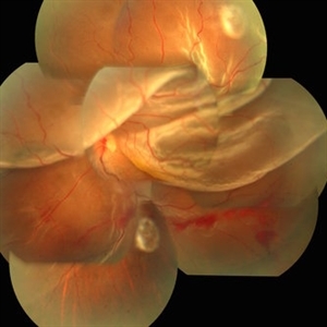
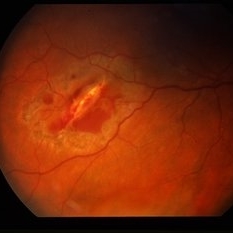
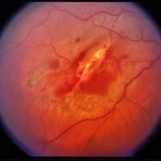
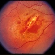
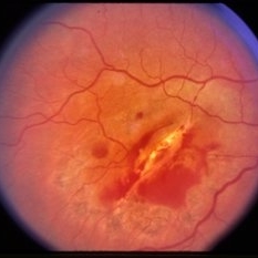
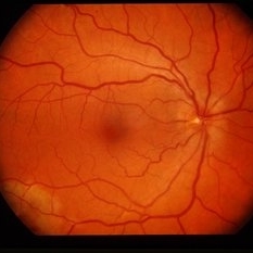
Patient presenting with globe perforation with a penetration seen at below the inferior arcade with some scattered hemorrhage and a retinal detachment.
Photographer: Rakesh Juneja
Condition/keywords: globe perforation
-
 Endophthalmitis
Jul 12 2014 by Yuntao Hu, MD, PhD
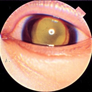
Eye picture of a six year-old boy with endophthalmitis in left eye after a tiny penetration.
Photographer: Yuntao Hu, Peking University Eye Center, Peking University Third Hospital
Condition/keywords: endophthalmitis
-
 Geographic Atrophy in Dry AMD
Dec 12 2019 by Darin R. Goldman, MD
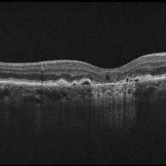
This OCT B-scan shows geographic atrophy (GA) in dry age-related macular degeneration. There is focal atrophy of the RPE and outer retinal layers underneath the fovea, which is typical of GA. The loss of RPE in the affected area, relative to the surrounding macula, results in reverse shadowing within the underlying choroid. This effect is due to more penetration of the optical signal from the OCT illumination source owing to a relative absence of light being reflected as it normally would be from intact RPE. The result is a distinct border on each side of the affected area, where the underlying choroidal signal is more intense than the immediately adjacent areas. Additionally, adjacent to the area of GA are typical drusen, which are nodule-like diffusely hyperreflective accumulations within and under the RPE/Bruch complex, and pigment epithelial detachments (PEDs), which are nodule-like elevations of the RPE with underlying hyporeflective spaces.
Condition/keywords: dry age-related macular degeneration (dry AMD), geographic atrophy, optical coherence tomography (OCT)
-
 Intraocular Foreign Body, Human Eye Lash, Post Trauma - Right Eye
Oct 3 2012 by James B. Soque, CRA, OCT-C, COA, FOPS
30-year-old white male, with nail gun misfire and blunt force trauma to right eye. Nail penerated sup-nas globe OD, at the equator. Force of nail penetration, 'brought in' an Eye Lash Folicle. During vitrectomy, sterile FB OD was noted. Photograph captured 1 week post op. Color Fundus Photo, 50 Deg, Max 1X
Photographer: James Soque, CRA, COA, Island Retina, Shirley, NY, USA
Imaging device: Topcon TRC 50 DX, OIS Imaging Software
Condition/keywords: blunt trauma, intraocular foreign body, penetration
-
 Lignocaine Retinal Toxicity
Aug 21 2015 by Mallika Goyal, MD
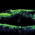
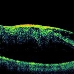
Right eye OCT of a 70-year-old male 3 weeks after inadvertent globe penetration with peribulbar anaesthesia needle and intraocular injection of lignocaine. There is a thick taut epimacular membrane with severely increased central retinal thickness. Fluorescein angiography revealed an occluded retinal arteriole at the macula indicating macular ischaemia underlying the membrane.
Photographer: Mallika Goyal, MD, Apollo Health City, Jubilee Hills, Hyderabad, India
Condition/keywords: lignocaine retinal toxicity
-
 Lignocaine Retinal Toxicity
Aug 21 2015 by Mallika Goyal, MD
Right eye OCT of a 70-year-old male 3 weeks after inadvertent globe penetration with peribulbar anaesthesia needle and intraocular injection of lignocaine shows a thick taut epimacular membrane with severely increased central retinal thickness. Fluorescein angiography revealed an occluded retinal arteriole at the macula indicating macular ischaemia underlying the membrane.
Photographer: Mallika Goyal, MD, Apollo Health City, Jubilee Hills, Hyderabad, India
Condition/keywords: lignocaine retinal toxicity
-
 Lignocaine Retinal Toxicity
Aug 21 2015 by Mallika Goyal, MD
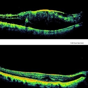
Right eye OCT of a 70-year-old male 3 weeks after inadvertent globe penetration with peribulbar anaesthesia needle and intraocular injection of lignocaine showing a taut epimacular membrane with macular elevation compared to a relatively normal foveal contour immediately after surgery suggesting progressive traction secondary to lignaocaine toxicity. Fluorescein angiography revealed an occluded retinal arteriole at the macula indicating macular ischaemia underlying the membrane.
Photographer: Mallika Goyal, MD, Apollo Health City, Jubilee Hills, Hyderabad, India
Condition/keywords: lignocaine retinal toxicity
-
 Lignocaine Retinal Toxicity
Aug 18 2015 by Mallika Goyal, MD
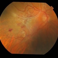
Right eye fundus of a 70-year-old male 3 weeks after inadvertent globe penetration with peribulbar anaesthesia needle and intraocular injection of lignocaine. There is a thick taut epimacular membrane with severely increased central retinal thickness. Fluorescein angiography revealed an occluded retinal arteriole at the macula indicating macular ischaemia underlying the membrane.
Photographer: Mallika Goyal, MD, Apollo Health City, Jubilee Hills, Hyderabad
Condition/keywords: lignocaine retinal toxicity
-
 Lignocaine Retinal Toxicity
Aug 18 2015 by Mallika Goyal, MD
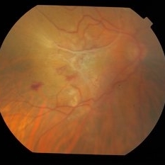
Right eye fundus of a 70-year-old male 3 weeks after inadvertent globe penetration with peribulbar anaesthesia needle and intraocular injection of lignocaine. There is a thick taut epimacular membrane with severely increased central retinal thickness. Fluorescein angiography revealed an occluded retinal arteriole at the macula indicating macular ischaemia underlying the membrane.
Photographer: Mallika Goyal, MD, Apollo Health City, Jubilee Hills, Hyderabad
Condition/keywords: lignocaine retinal toxicity
-
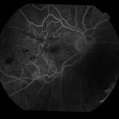 Lignocaine Retinal Toxicity
Aug 18 2015 by Mallika Goyal, MD
Right eye fluorescein angiogram of a 70-year-old male 3 weeks after inadvertent globe penetration with peribulbar anaesthesia needle and intraocular injection of lignocaine. There is an occluded retinal arteriole indicating macular ischaemia underlying the clinically obvious epimacular membrane.
Photographer: Mallika Goyal, MD, Apollo Health City, Jubilee Hills, Hyderabad
Condition/keywords: lignocaine retinal toxicity
-
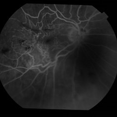 Lignocaine Retinal Toxicity
Aug 18 2015 by Mallika Goyal, MD
Right eye fluorescein angiogram of a 70-year-old male 3 weeks after inadvertent globe penetration with peribulbar anaesthesia needle and intraocular injection of lignocaine. There is an occluded retinal arteriole indicating macular ischaemia underlying the clinically obvious epimacular membrane.
Photographer: Mallika Goyal, MD, Apollo Health City, Jubilee Hills, Hyderabad
Condition/keywords: lignocaine retinal toxicity
-
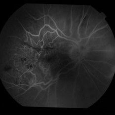 Lignocaine Retinal Toxicity
Aug 18 2015 by Mallika Goyal, MD
Right eye fluorescein angiogram of a 70-year-old male 3 weeks after inadvertent globe penetration with peribulbar anaesthesia needle and intraocular injection of lignocaine. There is an occluded retinal arteriole indicating macular ischaemia underlying the clinically obvious epimacular membrane.
Photographer: Mallika Goyal, MD, Apollo Health City, Jubilee Hills, Hyderabad
Condition/keywords: lignocaine retinal toxicity
-
 Penetrating Globe Injury
Oct 2 2013 by Jerald A. Bovino, MD
There was a metallic foreign body that caused a rutpured glove. An adjacent vitreous hemorrhage with posterior scleral rupture and adjacent choroidal infarction is present.
Condition/keywords: penetration, ruptured globe, vitreous hemorrhage
-
---thumb.jpg/image-square;max$300,300.ImageHandler) Penetrating Injury
Penetrating Injury
Oct 14 2013 by Maurice F. Rabb
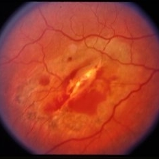
37 year old white male (hit by rock while mowing lawn) penetrating injury OD. This presentation is a 1-year follow-up on the case. Management of this case id becoming more complicated. We may well need more than just periocular steroids and laser treatment.
Condition/keywords: penetration
-
---thumb.jpg/image-square;max$300,300.ImageHandler) Penetrating Injury
Penetrating Injury
Oct 14 2013 by Maurice F. Rabb
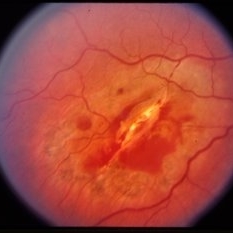
37 year old white male (hit by rock while mowing lawn) penetrating injury OD. This presentation is a 1-year follow-up on the case. Management of this case id becoming more complicated. We may well need more than just periocular steroids and laser treatment.
Condition/keywords: penetration
-
---thumb.jpg/image-square;max$300,300.ImageHandler) Penetrating Injury
Penetrating Injury
Oct 14 2013 by Maurice F. Rabb
37 year old white male (hit by rock while mowing lawn) penetrating injury OD. This presentation is a 1-year follow-up on the case. Management of this case id becoming more complicated. We may well need more than just periocular steroids and laser treatment.
Condition/keywords: penetration
-
 Penetrating ocular injury with posterior impact
Dec 19 2012 by Eric A. Postel, MD
Color fundus photo of a young make s/p penetrating injury with posterior segment impact and associated hemorrhage and commotio retinae
Condition/keywords: Berlin's edema, penetration, subretinal hemorrhage
-
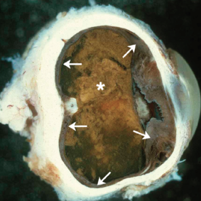 Sympathetic Ophthalmia
May 18 2020 by McGill University Health Centre
Sympathetic ophthalmia is characterized by bilateral diffuse granulomatous uveitis that occurs 2 weeks to many years after traumatic penetration or perforation of the eye. It threatens the sight of the uninjured (sympathizing) eye. In this enucleation specimen, thickening of the uveal tract is evident (arrows). Complete proteinaceous retinal detachment (*) is also present, along with posterior synechia (adhesion of the iris to the anterior capsule of the lens).
Condition/keywords: enucleation, sympathetic ophthalmia
-
 Trauma – Posterior Penetration
Aug 7 2015 by H. Michael Lambert, MD
Trauma – posterior penetration – no history, probably double perforation. Some laser surround the lesion. Fundus photo, inferotemporally OD.
Condition/keywords: laser, posterior perforation, trauma
-
 Trauma – Posterior Penetration
Aug 7 2015 by H. Michael Lambert, MD
Trauma – posterior penetration – no history, probably double perforation. Some laser surround the lesion. Fundus photo, inferotemporally OD.
Condition/keywords: laser, posterior perforation, trauma
-
 Trauma – Posterior Penetration
Aug 7 2015 by H. Michael Lambert, MD
Trauma – posterior penetration – no history, probably double perforation. Some laser surround the lesion. Fundus photo, inferotemporally OD.
Condition/keywords: laser, posterior perforation, trauma
-
 Trauma – Posterior Penetration
Aug 7 2015 by H. Michael Lambert, MD
Trauma – posterior penetration – no history, probably double perforation. Some laser surround the lesion. Fundus photo, inferotemporally OD.
Condition/keywords: laser, posterior perforation, trauma
-
 Trauma – Posterior Penetration
Aug 7 2015 by H. Michael Lambert, MD
Trauma – posterior penetration – no history, probably double perforation. Some laser surround the lesion. Fundus photo, inferotemporally OD.
Condition/keywords: laser, posterior perforation, trauma
-
 Trauma – Posterior Penetration
Aug 7 2015 by H. Michael Lambert, MD
Trauma – posterior penetration – no history, probably double perforation. Some laser surround the lesion. Fundus photo, inferotemporally OD.
Condition/keywords: laser, posterior perforation, trauma
-
 Trauma – Posterior Penetration
Aug 7 2015 by H. Michael Lambert, MD
Trauma – posterior penetration – no history, probably double perforation. Some laser surround the lesion. Fundus photo, inferotemporally OD.
Condition/keywords: laser, posterior perforation, trauma
 Loading…