Search results (98 results)
-
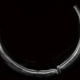 Wide-Field-OCT-montage
Jan 8 2018 by Netan Choudhry, MD, FRCS(C) FASRS
This is an SD-OCT montage image of a 55 year old male with optic neuropathy representing a wide-field OCT spanning 130 degrees.
Photographer: John Golding, Vitreous Retina Macula Specialists of Toronto
Imaging device: Heidelberg Spectralis OCT system
Condition/keywords: wide angle imaging
-
 Venous Loop
Feb 20 2024 by Soobien Lee
A 77-year-old male with a history of bilateral optic neuropathy from bilateral optic nerve sheath meningiomas S/P radiation/proton-beam therapies. Presented with radiation retinopathy OS and a known venous loop OS.
Photographer: Gavin Bragdon, Elman Retina Group
Imaging device: Optos Ultra-Widefield Imaging
Condition/keywords: Optos, OPTOS CALIFORNIA, radiation retinopathy, retinal vascular disease, venous loop
-
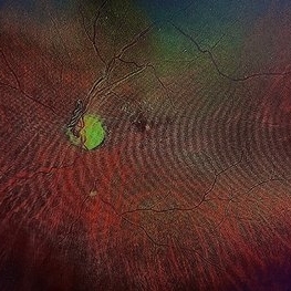
 Venous Loop
Feb 20 2024 by Soobien Lee
A 77-year-old male with a history of bilateral optic neuropathy from bilateral optic nerve sheath meningiomas S/P radiation/proton-beam therapies. Presented with radiation retinopathy OS and a known venous loop OS.
Photographer: Gavin Bragdon, Elman Retina Group
Imaging device: Optos Ultra-Widefield Fluorescein Angiography
Condition/keywords: fluorescein angiogram (FA), Optos, radiation retinopathy, retinal vascular disease, venous loop
-
 AION With Ciliotretinal Artery Occlusion
May 2 2013 by Henry J. Kaplan, MD
AION accompanied by partial CRAO which is visible as retinal edema and cherry red spot.
Condition/keywords: anterior ischemic optic neuropathy, central retinal artery occlusion (CRAO)
-
 Leukemic optic neuropathy
Oct 28 2022 by pedro fernandes souza neto
Fundus photograph of an 18-year-old woman with Leukemic optic neuropathy.
Photographer: Pedro Fernandes, Universidade Federal da Bahia
Condition/keywords: Leukemic optic neuropathy
-
 Hypertensive optic neuropathy and choroidopathy right eye
Jan 11 2013 by Alex P. Hunyor, MD
Previous hypertensive optic neuropathy and choroidopathy, right eye. A young female who had a history severe pre-eclampsia. Note optic atrophy and multiple Elschnig spots.
Condition/keywords: hypertensive choroidopathy, hypertensive optic neuropathy
-
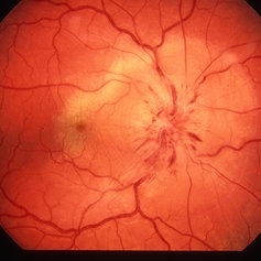
 Pseudo Foster Kennedy Syndrome
Oct 13 2022 by Aditya S Kelkar, MS, FRCS, FASRS,FRCOphth
Colour fundus photograph of a 44-year-old man showing bilateral small discs with optic atrophy on the right eye and disc edema on the left eye resulting from consecutive NAAION in both eyes.
Photographer: Dr Sukanya Mondal, National Institute of Ophthalmology, Pune. India
Imaging device: Zeiss Clarus 500
Condition/keywords: ischemic optic neuropathy, optic atrophy, optic disc edema
-
 Anterior Ischemic Optic Neuropathy and Choroidal Ischemia
Mar 1 2014 by Homayoun Tabandeh, MD, FASRS
Arteritic anterior ischemic optic neuropathy and choroidal ischemia in a patient with giant cell arteritis.
Condition/keywords: anterior ischemic optic neuropathy, giant cell arteritis
-
 Anterior Ischemic Optic Neuropathy and Choroidal Ischemia
Mar 1 2014 by Homayoun Tabandeh, MD, FASRS
Fundus fluorescein angiogram of a patient with arteritic anterior ischemic optic neuropathy and choroidal ischemia associated with giant cell arteritis.
Condition/keywords: anterior ischemic optic neuropathy
-
---thumb.JPG/image-square;max$300,300.ImageHandler) Traumatic Optic Neuropathy
Traumatic Optic Neuropathy
Nov 28 2012 by Mallika Goyal, MD
Left eye of a 19-year-old boy 4 weeks following a road accident involving head injury as chest compression with lung contusion has visual acuity CF CF. There is disc pallor with surrounding retinal edema and hemorrhages suggestive of traumatic optic neuropathy.
Photographer: Mallika Goyal, MD, Apollo Health City, Hyderabad, India
Condition/keywords: traumatic optic neuropathy
-
 AION
AION
Dec 19 2012 by Eric A. Postel, MD
fundus photograph of an elderly gentleman with AION
Condition/keywords: anterior ischemic optic neuropathy
-
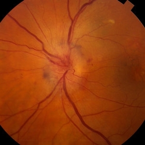
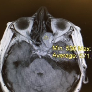
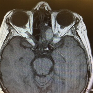 Acute Compressive Optic Neuropathy
Jun 1 2019 by John S. King, MD
84-year-old white female with acute loss of vision in the left eye one day ago was sent here after going to the ED per primary eye provider. She described vision loss as a grey curtain that became total darkness. She had left sided temporal tenderness and some left sided neck pain. In the ED the cardiac work-up was u/r, the ESR and CRP were normal, and the CTH showed some non-specific opacification in the L ethmoid sinus. Acuity was HM OS with RAPD, normal EOMs, no proptosis or ptosis, posteriorly no SVPs were noted; the optic discs were pink and flat; no emboli or retinal whitening present; some bear tracks located nasally (see photo). She was referred to Dr. Doyle, who ordered an MRI, which showed a large mucocele with bony erosion into the left orbit, along with some ON enhancement possibly from compression (see images). She was operated that night and later recovered to 20/40 in that eye with a residual, inferior arcuate scotoma.
Condition/keywords: bear tracks, optic neuropathy
-
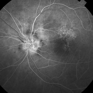
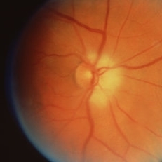
 Acute Optic Neuropathy Due to Large Mucocele
Jun 1 2019 by John S. King, MD
84-year-old white female with acute loss of vision in the left eye one day ago was sent here after going to the ED per primary eye provider. She described vision loss as a grey curtain that became total darkness. She had left sided temporal tenderness and some left sided neck pain. In the ED the cardiac work-up was u/r, the ESR and CRP were normal, and the CTH showed some non-specific opacification in the L ethmoid sinus. Acuity was HM OS with RAPD, normal EOMs, no proptosis or ptosis, posteriorly no SVPs were noted; the optic discs were pink and flat; no emboli or retinal whitening present; some bear tracks located nasally (see photo). She was referred to Dr. Doyle, who ordered an MRI, which showed a large mucocele with bony erosion into the left orbit, along with some ON enhancement possibly from compression (see Images). She was operated that night and later recovered to 20/40 in that eye with a residual, inferior arcuate scotoma.
Condition/keywords: bear tracks, optic neuropathy
-
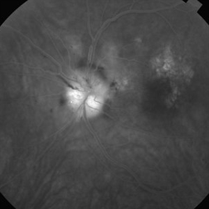
 Acute Optic Neuropathy Due to Large Mucocele (Incidental Bear Tracks)
Jun 1 2019 by John S. King, MD
84-year-old white female with acute loss of vision in the left eye one day ago was sent here after going to the ED per primary eye provider. She described vision loss as a grey curtain that became total darkness. She had left sided temporal tenderness and some left sided neck pain. In the ED the cardiac work-up was u/r, the ESR and CRP were normal, and the CTH showed some non-specific opacification in the L ethmoid sinus. Acuity was HM OS with RAPD, normal EOMs, no proptosis or ptosis, posteriorly no SVPs were noted; the optic discs were pink and flat; no emboli or retinal whitening present; some bear tracks located nasally (see photo). She was referred to Dr. Doyle, who ordered an MRI, which showed a large mucocele with bony erosion into the left orbit, along with some ON enhancement possibly from compression (see images). She was operated that night and later recovered to 20/40 in that eye with a residual, inferior arcuate scotoma.
Photographer: Karin Aletter
Imaging device: Topcon 50
Condition/keywords: bear tracks, optic neuropathy
-
---thumb.JPG/image-square;max$300,300.ImageHandler) Anterior Ischaemic Optic Neuropathy
Anterior Ischaemic Optic Neuropathy
Nov 18 2013 by Mallika Goyal, MD
Second event of AION involving the lower half of otpic nerve in a patient with superior half optic atrophy from prior AION.
Photographer: Mallika Goyal, MD, Apollo Health City, Hyderabad
Condition/keywords: anterior ischemic optic neuropathy
-
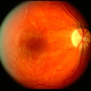
 Anterior Ischaemic Optic Neuropathy
Oct 25 2012 by Mallika Goyal, MD
Fundus photograph of the left eye of a 65-year-old diabetic gentleman with sudden vision drop 3 days prior to presentation shows pale disc edema.
Condition/keywords: anterior ischemic optic neuropathy
-
---thumb.JPG/image-square;max$300,300.ImageHandler) Anterior Ischaemic Optic Neuropathy
Anterior Ischaemic Optic Neuropathy
Nov 18 2013 by Mallika Goyal, MD
Unilateral inferior disc edema in a diabetic patient with superior altitudinal field loss suggestive of AION.
Photographer: Mallika Goyal, MD, Apollo Health City, Hyderabad
Condition/keywords: anterior ischemic optic neuropathy
-
---thumb.JPG/image-square;max$300,300.ImageHandler) Anterior Ischaemic Optic Neuropathy
Anterior Ischaemic Optic Neuropathy
Dec 7 2013 by Mallika Goyal, MD
Right eye of a diabetic 65-year-old gentleman with superior half optic disc pallor 2 months following sudden vision drop from anterior ischaemic optic neuropathy. Superior disc pallor corresponds to inferior altitudinal field defect. There was no visual or field improvement following oral steroids.
Photographer: Mallika Goyal, MD, Apollo Health City, Hyderabad, India
Condition/keywords: anterior ischemic optic neuropathy
-
 Anterior Ischemic Optic Neruopathy
Feb 20 2013 by From the Collections of Thomas M. Aaberg, MD and Thomas M. Aaberg Jr., MD
With atrophy and edema and small hemorrhage; 20/100.
Condition/keywords: anterior ischemic optic neuropathy
-
 Anterior Ischemic Optic Neruopathy
Feb 20 2013 by From the Collections of Thomas M. Aaberg, MD and Thomas M. Aaberg Jr., MD
With atrophy and disc edema.
Condition/keywords: anterior ischemic optic neuropathy
-
 Anterior Ischemic Optic Neruopathy
Feb 20 2013 by From the Collections of Thomas M. Aaberg, MD and Thomas M. Aaberg Jr., MD
Fluorescein of anterior ischemic optic neruopathy.
Condition/keywords: anterior ischemic optic neuropathy
-
 Anterior Ischemic Optic Neruopathy
Feb 20 2013 by From the Collections of Thomas M. Aaberg, MD and Thomas M. Aaberg Jr., MD
Fluorescein for anterior ischemic optic neruopathy.
Condition/keywords: anterior ischemic optic neuropathy
-
 Anterior Ischemic Optic Neuropathy
Mar 26 2019 by Gary R. Cook, MD, FACS
Anterior ischemic optic neuropathy OD.
Condition/keywords: anterior ischemic optic neuropathy
-
---thumb.jpg/image-square;max$300,300.ImageHandler) Anterior Ischemic Optic Neuropathy
Anterior Ischemic Optic Neuropathy
Mar 29 2013 by Henry J. Kaplan, MD
Anterior Ischemic Optic Neuropathy; notice the typical pale optic disc swelling and faint splinter hemorrhages.
Condition/keywords: anterior ischemic optic neuropathy
 Loading…