Search results (63 results)
-
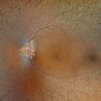
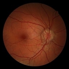
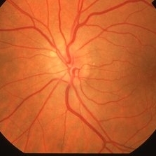
 Optic Nerve Head Drusen With Idiopathic CNV
Feb 17 2017 by Kristen Wagner
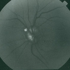
22-year-old female fundus photograph of a right eye with Optic Nerve Drusen with Idiopathic CNV.
Photographer: Kristen Wagner, COT, OSC Ophthalmic Photographer, Tennessee Retina, Nashville TN
Condition/keywords: choroidal neovascularization (CNV), drusen of optic disc, optic disc drusen
-
 Optic Disc Drusen, RP
Apr 21 2025 by Virginia Gebhart
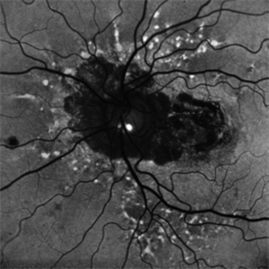
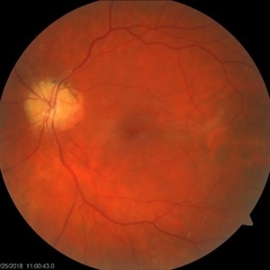
28 year old male with stable retinitis pigmentosa and optic disc drusen OU. Bardet-Biedl variant identified in previous genetic testing. BCVA 20/50 OD, 20/30 OS
Photographer: Virginia Gebhart, Retina Consultants of Carolina
Imaging device: Optos California
Condition/keywords: Drusen, optic disc drusen, retinitis pigmentosa
-
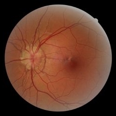
 Myelinated Nerve Fibers
Apr 18 2025 by DR Rohit Gupta
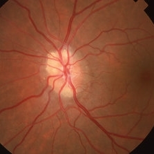
The **myelinated nerve fibers of the optic disc** (also known as **medullated nerve fibers**) are retinal nerve fibers that retain their myelin sheath as they pass through the optic nerve head. Normally, retinal nerve fibers are unmyelinated to allow for light transparency, but in some cases, myelination extends anteriorly into the retina, appearing as a striking white, feathery patch on the optic disc or peripapillary retina. ### **Key Features:** 1. **Appearance:** - Dense, white, striated patches with feathery edges. - Typically located at the superior or inferior pole of the optic disc. - May obscure retinal vessels underneath. 2. **Clinical Significance:** - Usually **benign** and asymptomatic. - **Congenital** (present at birth or early childhood). - Rarely associated with **visual field defects** (e.g., scotomas corresponding to the area of myelination). - Occasionally linked with **high myopia** or **amblyopia** if extensive. 3. **Pathophysiology:** - Failure of oligodendrocytes or Schwann cells to stop myelination at the lamina cribrosa. - Normally, myelination stops at the optic nerve head, but in this condition, it extends into the retina. 4. **Diagnosis:** - **Fundoscopy:** Classic white, feathery appearance. - **Optical Coherence Tomography (OCT):** Shows thickened retinal nerve fiber layer (RNFL). - **Visual Field Testing:** May detect defects if large. 5. **Differential Diagnosis:** - Optic disc edema - Cotton wool spots - Retinoblastoma (rarely, but must be ruled out in children) 6. **Management:** - No treatment required if asymptomatic. - Monitor for amblyopia in children. - Rare cases with significant visual impairment may need further evaluation. ### **Fun Fact:** Myelinated nerve fibers are seen in **~0.5-1%** of the population and are usually an incidental finding.
Photographer: Dr Rohit gupta
Imaging device: Samsung S21
Condition/keywords: Medulated Nerve fibre, Medullated Nerve fibres, myelinated nerve fibers, Myelinated Nerve Fibres, optic disc drusen
-
 Optic Disc Drusen
Jun 29 2022 by Mohamed Awadalla, MD, FRCSEd
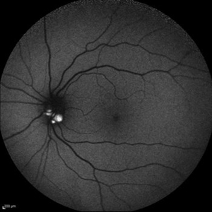
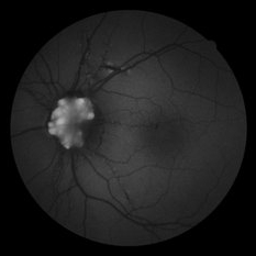
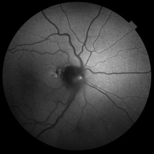
Autofluorescence in optic disc drusen Red free fundus photo
Condition/keywords: Autofluorescence, optic disc drusen
-
 Optic Disc Drusen
Jul 10 2013 by Hamid Ahmadieh, MD
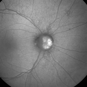
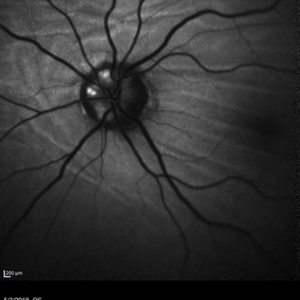
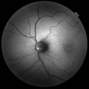
Fundus autofluorescence image of the left eye of a 24-year-old woman with optic disc drusen and VA 20/20.
Photographer: Solmaz Shahmohammad, Negah Eye Center, Tehran
Imaging device: Heidelberg Spectralis
Condition/keywords: fundus autofluorescence (FAF), optic disc drusen
-
 Optic disc drusen
Dec 25 2012 by Alex P. Hunyor, MD
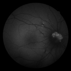
Autofluorescent image of the right optic disc showing autofluorescence of optic disc drusen.
Condition/keywords: optic disc drusen
-
 Optic Disc Drusen and Angioid Streaks
Jun 3 2020 by Mirko Ratkovic, MD
Optic disc drusen and angioid streaks.
Condition/keywords: angioid streaks, drusen of optic disc, fundus autofluorescence (FAF), fundus photograph
-
 Optic Disc Drusen and Angioid Streaks
Jun 3 2020 by Mirko Ratkovic, MD
Optic disc drusen and angioid streaks.
Condition/keywords: angioid streaks, fundus autofluorescence (FAF), fundus photograph, optic disc drusen
-
 Optic Disc Drusen and Angioid Streaks
Jun 3 2020 by Mirko Ratkovic, MD
Optic disc drusen and angioid streaks.
Condition/keywords: angioid streaks, fundus autofluorescence (FAF), fundus photograph, optic disc drusen
-
 Optic Disc Drusen and Angioid Streaks
Jun 3 2020 by Mirko Ratkovic, MD
Optic disc drusen and angioid streaks.
Condition/keywords: angioid streaks, fundus autofluorescence (FAF), optic disc drusen
-
 Optic Disc Drusen Autofluorescence
Jul 16 2023 by Aditya S Kelkar, MS, FRCS, FASRS,FRCOphth
Fundus autofluorescence imaging of a 22-year-old male with optic disc drusen seen as hyperautofluorescent spot.
Photographer: Dr. Harsh Jain
Condition/keywords: Autofluorescence imaging of Optic Disc Drusen
-
 Choroidal Folds and Optic Disc Drusen
Aug 1 2018 by Emily Cooper
Fundus autofluorescence photo of a 62-year-old man who presented for evaluation of choroidal folds and optic disc drusen. He is currently following up with neuro-ophthalmology and has suspected intracranial hypertension.
Photographer: Emily Cooper, Retina Specialists of Michigan
Condition/keywords: choroidal folds, drusen of optic disc
-
---thumb.jpg/image-square;max$300,300.ImageHandler) Optic Disc Drusen
Optic Disc Drusen
Mar 27 2013 by Henry J. Kaplan, MD
Autofluorescence imaging shows heper AF on the optic nerve head specially superiorly due to drusen in the same patient #2.
Imaging device: Heidelberg spectralis
Condition/keywords: drusen of optic disc, optic disc drusen, optic nerve drusen
-
 Optic Disc Drusen
Sep 21 2012 by Suber S. Huang, MD, MBA, FASRS
Optic disc drusen demonstrating autofluorescence
-
 AF of Disc Drusen
Mar 26 2019 by Gary R. Cook, MD, FACS
Autofluorescent (AF) image of the left eye of a 60-year-old with bilateral optic disc drusen; VA= 20/15.
Imaging device: Topcon VT-50
Condition/keywords: drusen of optic disc, optic disc drusen
-
 Angioid Streaks/Optic Disc Drusen
Oct 30 2024 by JULIAN VILLARREAL, MD
FAF showing angiod streaks , optic disc drusen, and macular atrophy secondary to macular neovascular membrane.
Photographer: Julián Villarreal MD
Imaging device: Mirante
Condition/keywords: Angioid Streaks, macular atrophy, optic disc drusen
-
 Autofluorescence in Optic Nerve Head Drusen
May 28 2024 by Nishikant J Borse, MS, FMRF, FASRS
65-year-old female was referred for disc edema. An Autofluorescence Imaging was done which showed the autofluorescence of the optic nerve head drusen.
Photographer: Dr Nishikant Borse , Insight eye Clinic , Mumbai
Imaging device: Topcon Triton
Condition/keywords: Autofluorescence imaging of Optic Disc Drusen
-
 Autofluorescence of Optic Disc Drusen
Mar 2 2014 by Homayoun Tabandeh, MD, FASRS
Autofluorescence of optic disc drusen.
Condition/keywords: optic disc drusen
-
 Buried drusen with CNV
Dec 19 2012 by Eric A. Postel, MD
Buried optic disc drusen complicated by peripapillary subretinal neovascularisation
Condition/keywords: choroidal neovascularization (CNV), optic disc drusen
-
 Color Fundus Photographs of Optic Disc Drusen
Apr 26 2018 by Ahmad B. Tarabishy, MD
Fundus photographs and autofluorescence of a 75-year-old man with an epiretinal membrane in the left eye. Incidentally, he had a history of optic disc drusen, which show a striking hyperautofluorescence on FAF imaging.
Photographer: Michelle Howarth, Lakeland Eye Clinic
Imaging device: Zeiss Visucam
Condition/keywords: fundus autofluorescence (FAF), optic disc drusen
-
 Drusen of Optic Nerve Head
Mar 26 2019 by Gary R. Cook, MD, FACS
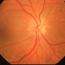
31-year-old Indian male with visible drusen of the optic nerve OS; VA= 20/20.
Imaging device: Topcon VT-50
Condition/keywords: drusen of optic disc, optic disc drusen
-
 Drusen of Optic Nerve Head
Mar 26 2019 by Gary R. Cook, MD, FACS
Right eye of a 60-year-old white male with visible drusen of the optic disc bilaterally; VA= 20/15.
Imaging device: Topcon VT-50
Condition/keywords: drusen of optic disc, optic disc drusen
-
 Drusen of Optic Nerve Head
Mar 26 2019 by Gary R. Cook, MD, FACS
Left eye of a 60-year-old white male with bilateral optic disc drusen; VA= 20/15.
Imaging device: Topcon VT-50
Condition/keywords: drusen of optic disc, optic disc drusen
 Loading…