Search results (70 results)
-
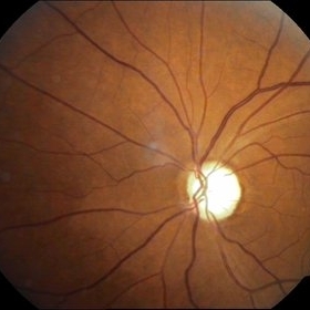
 Disseminated Chorioretinitis With Unknown Etiology
Apr 5 2018 by Kim Barrett
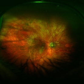
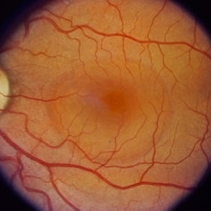
Ultra-wide field fluorescein angiogram of a 31-year-old female with intermittent pain in her left eye. Her condition has been managed in Liberia until recently when she moved to the United States. She suffers from multiple modalities including central retinal artery occlusion, posterior synechiae of the iris, interstitial keratitis, disseminated chorioretinitis, as well as HIV. An infectious cause is high on the differential in light of her HIV status. DDx: hypertensive crisis, an embolism (? IV drug use), coagulopathy, trauma, infectious. Blood work was normal. Her current vision is 20/30 right eye and 20/400 left eye.
Photographer: Kim Barrett, COA
Imaging device: Optos
Condition/keywords: central retinal artery occlusion (CRAO), chorioretinal scar, ciliary artery sparring, disseminated chorioretinitis, HIV, left eye, optic atrophy, staining
-
 Hypertensive optic neuropathy and choroidopathy right eye
Jan 11 2013 by Alex P. Hunyor, MD
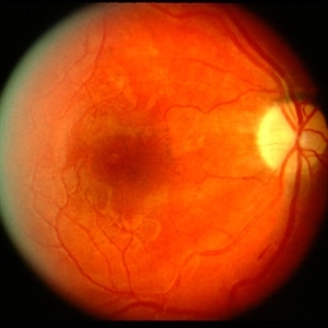
Previous hypertensive optic neuropathy and choroidopathy, right eye. A young female who had a history severe pre-eclampsia. Note optic atrophy and multiple Elschnig spots.
Condition/keywords: hypertensive choroidopathy, hypertensive optic neuropathy
-
 Optic Atrophy and Attenuated Retinal Vessels Following Endophthalmitis
Jul 12 2014 by Philip J. Polkinghorne, MD
This elderly lady underwent a vitrectomy for post-surgical endophthalmitis. The infection was successfully treated but the functional outcome was poor because of optic atrophy and attenuated retinal vessels.
Photographer: Alex Fraser
Imaging device: Optos Camera
Condition/keywords: attenuated vessels, endophthalmitis, optic atrophy, post-vitrectomy
-
 Pseudo Foster Kennedy Syndrome
Oct 13 2022 by Aditya S Kelkar, MS, FRCS, FASRS,FRCOphth
Colour fundus photograph of a 44-year-old man showing bilateral small discs with optic atrophy on the right eye and disc edema on the left eye resulting from consecutive NAAION in both eyes.
Photographer: Dr Sukanya Mondal, National Institute of Ophthalmology, Pune. India
Imaging device: Zeiss Clarus 500
Condition/keywords: ischemic optic neuropathy, optic atrophy, optic disc edema
-
 Syphilis Neuroretinopathy
Apr 2 2018 by JEFFERSON R SOUSA, Tecg.º (Biomedical Systems Technology)
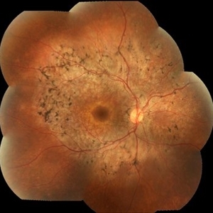
Female patient, 21-years-old, with complaint of low vision in the right eye for 3 years. According to information from the patient's history, at the time she noticed the low vision, it also coincided with a picture of a strong urinary infection as well as episodes of constant tonsillitis. Yes, the patient did not seek medical attention and self-medicated with antibiotics. In ophthalmologic evaluation, as well as examinations of color retinography and ocular fundus autofluorescence, important pigmentary alterations were observed following vascular arches with pigment mobilization in osteoclasts (aspect of a unilateral pigmentary retinitis secondary to the inflammatory process). Which suggested inflammatory process sequelae. Through the laboratory tests, he had positive (+) confirmation for SYPHILIS NEURORETINOPATHY .
Photographer: JEFFERSON R SOUSA - Study Center and Ophthalmological Research Dr. Andre M V Gomes, Institute Dr. Suel Abujamra São Paulo-Brazil
Imaging device: Fundus camera Topcon TRC-50 DX, Imaginet 5.0, angle de 50 graus. Flash 36 / Mosaic with 10 images.
Condition/keywords: neurosyphilitic optic atrophy, retinitis pigmentosa, syphilis, syphilis neuroretinopathy
-
 Wyburn Mason Racemose Angiomatosis
May 22 2016 by Olivia Rainey
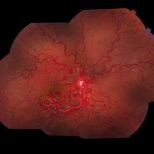
Color fundus montage of an 13-year-old female with arteriovenous malformation (Wyburn Mason Racemose Angiomatosis) affecting her right eye. The retinal arteriovenous malformation appears to be stable. She presented with NLP in the eye, strabismus, and peripheral retinal ischemia. She is at risk for neovascular complications; however, she is currently being treated with Sirolimus. Since she is on this systemically, there is no need to perform intraocular anti-VEGF injections or PRP laser. She also presented with optic atrophy affecting her left eye, secondary to chiasmal involvement of arteriovenous malformation. She has had a potential progressive visual field loss involving the temporal aspect of her visual field from the left eye. There is sector optic atrophy. Presumably, this is due to a compressive effect of her arteriovenous malformation on the nasal nerve fiber layer (corresponding to the temporal visual field) crossing to the right occipital cortex at the chiasm.
Photographer: Olivia Rainey
Imaging device: Topcon 50dx
Condition/keywords: arteriovenous malformation, color fundus photograph, color photo, montage, peripheral ischemia, Sirolimus
-
 Glaucomatous Optic Atrophy
Sep 20 2014 by Mehul A Shah
A 65-year-old male presented with loss of vision and found to have glaucomatous optic atrophy.
Photographer: Drashti Netralaya,Dahod
Imaging device: Zeiss ff450
Condition/keywords: glaucomatous atrophy of optic disc
-
---thumb.JPG/image-square;max$300,300.ImageHandler) Anterior Ischaemic Optic Neuropathy
Anterior Ischaemic Optic Neuropathy
Nov 18 2013 by Mallika Goyal, MD
Second event of AION involving the lower half of otpic nerve in a patient with superior half optic atrophy from prior AION.
Photographer: Mallika Goyal, MD, Apollo Health City, Hyderabad
Condition/keywords: anterior ischemic optic neuropathy
-
---thumb.jpg/image-square;max$300,300.ImageHandler) Behcet Disease
Behcet Disease
Feb 15 2013 by From the Collections of Thomas M. Aaberg, MD and Thomas M. Aaberg Jr., MD
Reprint of a fundus photograph from a patient with Behcet disease showing optic atrophy, vessel narrowing, and pigmentary changes (from Colvard et al, Arch Ophthalmol 1977;95(10):1813-7).
Condition/keywords: posterior uveitis, retinitis
-
 Bilateral Calcific Retina Arteriolar Occlusions in a Patient with Metastatic Ovarian Carcinoma
Dec 10 2020 by McGill University Health Centre
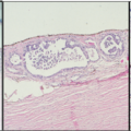
47-year-old female with cough and fever. Imaging showed a right pulmonary infiltrate. Transbronchial needle biopsy revealed lymphangitic spread of papillary adenocarcinoma with psammoma bodies (MRI of thyroid, CT of abdomen and pelvis were negative) gynecologic evaluation negative at that time . The patient had bilateral floaters, VA: 20/40 OD and 20/20 OS. Fundus examination showed retinal arteriolar sheathing and a flat choroidal lesion OS and vitritis OD. Fluorescein angiogram showed staining of left superior temporal retinal arterioles and bilateral midperipheral patchy hyperfluorescence at RPE. The patient vision in the OD deteriorated to 20/400, and in the OS 20/50. Four months later a new choroidal lesion was diagnosed OS. An abdominal mass consistent with a cystadenoma of the ovary was diagnosed. After a year patient developed systemic metastasis. Autopsy: Metastatic adenocarcinoma to the lung, both adrenals, para-aortic lymph nodes, left hip, right breast, occipital skin, serosal surface of liver, pituitary. In almost all metastatic lesions psammoma bodies were found. Presumptive diagnosis is a primary tumor of the ovary. Histopathologic examination of both eyes disclosed : Bilateral metastatic adenocarcinoma to the vitreous with partially calcified proliferation along internal limiting membrane, OS. Metastatic adenocarcinoma to choroid, OS. Bilateral optic atrophy secondary to retinal arteriolar occlusion with calcification.
Condition/keywords: bilateral, calcification, histopathology, metastatic adenocarcinoma, pathology, retinal arteriolar occlusion
-
 Bilateral Calcific Retina Arteriolar Occlusions in a Patient with Metastatic Ovarian Carcinoma
Dec 10 2020 by McGill University Health Centre
47-year-old female with cough and fever. Imaging showed a right pulmonary infiltrate. Transbronchial needle biopsy revealed lymphangitic spread of papillary adenocarcinoma with psammoma bodies (MRI of thyroid, CT of abdomen and pelvis were negative) gynecologic evaluation negative at that time . The patient had bilateral floaters, VA: 20/40 OD and 20/20 OS. Fundus examination showed retinal arteriolar sheathing and a flat choroidal lesion OS and vitritis OD. Fluorescein angiogram showed staining of left superior temporal retinal arterioles and bilateral midperipheral patchy hyperfluorescence at RPE The patient vision in the OD deteriorated to 20/400, and in the OS 20/50. Four months later a new choroidal lesion was diagnosed OS. An abdominal mass consistent with a cystadenoma of the ovary was diagnosed. After a year patient developed systemic metastasis. Autopsy: Metastatic adenocarcinoma to the lung, both adrenals, para-aortic lymph nodes, left hip, right breast, occipital skin, serosal surface of liver, pituitary. In almost all metastatic lesions psammoma bodies were found. Presumptive diagnosis is a primary tumor of the ovary. Histopathologic examination of both eyes disclosed : Bilateral metastatic adenocarcinoma to the vitreous with partially calcified proliferation along internal limiting membrane, OS. Metastatic adenocarcinoma to choroid, OS. Bilateral optic atrophy secondary to retinal arteriolar occlusion with calcification.
Condition/keywords: bilateral, calcification, histopathology, metastatic adenocarcinoma, pathology, retinal arteriolar occlusion
-
 Bilateral Calcific Retina Arteriolar Occlusions in a Patient with Metastatic Ovarian Carcinoma
Dec 10 2020 by McGill University Health Centre
47-year-old female with cough and fever. Imaging showed a right pulmonary infiltrate. Transbronchial needle biopsy revealed lymphangitic spread of papillary adenocarcinoma with psammoma bodies (MRI of thyroid, CT of abdomen and pelvis were negative) gynecologic evaluation negative at that time . The patient had bilateral floaters, VA: 20/40 OD and 20/20 OS. Fundus examination showed retinal arteriolar sheathing and a flat choroidal lesion OS and vitritis OD. Fluorescein angiogram showed staining of left superior temporal retinal arterioles and bilateral midperipheral patchy hyperfluorescence at RPE. The patient vision in the OD deteriorated to 20/400, and in the OS 20/50. Four months later a new choroidal lesion was diagnosed OS. An abdominal mass consistent with a cystadenoma of the ovary was diagnosed. After a year patient developed systemic metastasis. Autopsy: Metastatic adenocarcinoma to the lung, both adrenals, para-aortic lymph nodes, left hip, right breast, occipital skin, serosal surface of liver, pituitary. In almost all metastatic lesions psammoma bodies were found. Presumptive diagnosis is a primary tumor of the ovary. Histopathologic examination of both eyes disclosed : Bilateral metastatic adenocarcinoma to the vitreous with partially calcified proliferation along internal limiting membrane, OS. Metastatic adenocarcinoma to choroid, OS. Bilateral optic atrophy secondary to retinal arteriolar occlusion with calcification.
Condition/keywords: bilateral, calcification, histopathology, metastatic adenocarcinoma, pathology, retinal arteriolar occlusion
-
 Bilateral Calcific Retina Arteriolar Occlusions in a Patient with Metastatic Ovarian Carcinoma
Dec 10 2020 by McGill University Health Centre
47-year-old female with cough and fever. Imaging showed a right pulmonary infiltrate. Transbronchial needle biopsy revealed lymphangitic spread of papillary adenocarcinoma with psammoma bodies (MRI of thyroid, CT of abdomen and pelvis were negative) gynecologic evaluation negative at that time Patient had bilateral floaters, VA: 20/40 OD and 20/20 OS. Fundus examination showed retinal arteriolar sheathing and a flat choroidal lesion OS and vitritis OD. Fluorescein angiogram showed staining of left superior temporal retinal arterioles and bilateral midperipheral patchy hyperfluorescence at RPE The patient vision in the OD deteriorated to 20/400, and in the OS 20/50. Four months later a new choroidal lesion was diagnosed OS. An abdominal mass consistent with a cystadenoma of the ovary was diagnosed. After a year patient developed systemic metastasis. Autopsy: Metastatic adenocarcinoma to the lung, both adrenals, para-aortic lymph nodes, left hip, right breast, occipital skin, serosal surface of liver, pituitary. In almost all metastatic lesions psammoma bodies were found. Presumptive diagnosis is a primary tumor of the ovary. Histopathologic examination of both eyes disclosed : Bilateral metastatic adenocarcinoma to the vitreous with partially calcified proliferation along internal limiting membrane, OS. Metastatic adenocarcinoma to choroid, OS. Bilateral optic atrophy secondary to retinal arteriolar occlusion with calcification.
Condition/keywords: bilateral, calcification, histopathology, metastatic adenocarcinoma, pathology, retinal arteriolar occlusion
-
 Bilateral Calcific Retina Arteriolar Occlusions in a Patient with Metastatic Ovarian Carcinoma
Dec 10 2020 by McGill University Health Centre
47-year-old female with cough and fever. Imaging showed a right pulmonary infiltrate. Transbronchial needle biopsy revealed lymphangitic spread of papillary adenocarcinoma with psammoma bodies (MRI of thyroid, CT of abdomen and pelvis were negative) gynecologic evaluation negative at that time . The patient had bilateral floaters, VA: 20/40 OD and 20/20 OS. Fundus examination showed retinal arteriolar sheathing and a flat choroidal lesion OS and vitritis OD. Fluorescein angiogram showed staining of left superior temporal retinal arterioles and bilateral midperipheral patchy hyperfluorescence at RPE The patient vision in the OD deteriorated to 20/400, and in the OS 20/50. Four months later a new choroidal lesion was diagnosed OS. An abdominal mass consistent with a cystadenoma of the ovary was diagnosed. After a year patient developed systemic metastasis. Autopsy: Metastatic adenocarcinoma to the lung, both adrenals, para-aortic lymph nodes, left hip, right breast, occipital skin, serosal surface of liver, pituitary. In almost all metastatic lesions psammoma bodies were found. Presumptive diagnosis is a primary tumor of the ovary. Histopathologic examination of both eyes disclosed : Bilateral metastatic adenocarcinoma to the vitreous with partially calcified proliferation along internal limiting membrane, OS. Metastatic adenocarcinoma to choroid, OS. Bilateral optic atrophy secondary to retinal arteriolar occlusion with calcification.
Condition/keywords: bilateral, calcification, histopathology, metastatic adenocarcinoma, pathology, retinal arteriolar occlusion
-
 BRVO With Optic Atrophy
Nov 27 2020 by Sham Talati, DOMS
A 55-year-old male patient presented with glaucomatous optic atrophy with ST BRVO in the right eye.
Photographer: Dr. Sham Talati,Retina Foundation,Ahmedabad
Imaging device: Nidek Mirante
Condition/keywords: branch retinal vein occlusion (BRVO), branch vein occlusion (BVO), glaucomatous atrophy of optic disc, optic atrophy
-
 Central Serous Chorioretinopathy (CSR)
Sep 21 2023 by Ben Serar
Fundus photograph showing increased cup-disc ratio with nasalisation of vessels , with thinning of Neuroretinal rim and bayonetting of vessels in a case of Glaucomatous Optic Atrophy (GOA) Fundus photograph of LE showing serous macular detachment in a case of Central Serous Chorioretinopathy (CSR).
Condition/keywords: Central Serous Chorioretinopathy (CSR)
-
 Choroidal Melanoma Kissing the Disc and Macula
Oct 5 2018 by Nishikant J Borse, MS, FMRF, FASRS
Choroidal melanoma in a 42-year-old male. Unfortunately this left eye is the only good eye. The right eye has optic atrophy.
Photographer: Dr Nishikant borse
Imaging device: Kowa
-
 Choroidal Melanoma Kissing the Disc and Macula
Oct 5 2018 by Nishikant J Borse, MS, FMRF, FASRS
Choroidal melanoma in a 42-year-old male. Unfortunately this left eye is the only good eye. The right eye has optic atrophy.
Photographer: Dr Nishikant borse
Imaging device: Kowa
-
 Dengue Retinitis Healed With Optic Atrophy
Jul 29 2014 by Mallika Goyal, MD
Right fundus of a 53-year-old lady treated earlier with oral steroids for dengue fever and associated retinitis shows marked disc pallor and extensive RPE atrophy also involving the macula. Her vision improved from presentation after treatment with oral steroids.
Photographer: Mallika Goyal, MD, Apollo Health City, Jubilee Hills, Hyderabad-500033
Condition/keywords: Dengue retinitis
-
 Dengue Retinitis Healed With Optic Atrophy
Jul 29 2014 by Mallika Goyal, MD
Left fundus of a 53-year-old lady treated earlier with oral steroids for dengue fever and associated retinitis shows marked disc pallor and extensive RPE atrophy also involving the macula. Her vision improved from presentation after treatment with oral steroids.
Photographer: Mallika Goyal, MD, Apollo Health City, Jubilee Hills, Hyderabad-500033
Condition/keywords: Dengue retinitis
-
 Extensive/ Heavy Focal/ PRP OD - Color
Jun 28 2018 by Hosam Attia, MD
70-year-old woman, seen for initial eye exam, with endstage PDR and H/O prior Focal/ PRP OU , somewhere else.
Imaging device: Optos - California
Condition/keywords: chorioretinal scar, focal laser, ghost vessels, optic atrophy, pan-retinal photocoagulation (PRP), proliferative diabetic retinopathy (PDR)
-
 Extensive/ Heavy Focal/ PRP OD - FAF
Jun 28 2018 by Hosam Attia, MD
70-year-old woman, seen for initial eye exam, with endstage PDR and H/O prior Focal/ PRP OU , somewhere else.
Imaging device: Optos - California
Condition/keywords: chorioretinal scar, focal laser, ghost vessels, optic atrophy, pan-retinal photocoagulation (PRP), proliferative diabetic retinopathy (PDR)
 Loading…