Search results (30 results)
-
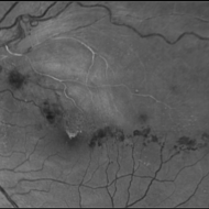
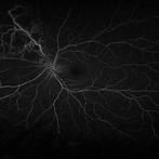
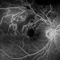 Branch Retinal Vein Occlusion- Fluorescein Angiogram, Montage
Apr 15 2016 by James B. Soque, CRA, OCT-C, COA, FOPS
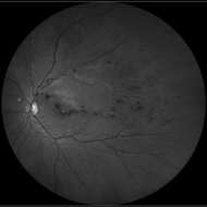
A fluorescein angiogram of an 80-year-old white female with a superotemporal branch retinal vein occlusion, and retinal edema of the right eye. Currently receiving Lucentis 0.5 injection therapy.
Photographer: James Soque, CRA OCT-C COA, Island Retina, Shirley, NY
Imaging device: Topcon TRC, MERGE Imaging Software V. 11.2.0
Condition/keywords: branch retinal vein occlusion (BRVO), montage, non-perfused branch retinal vein occlusion (BRVO)
-
 Vein Occlusion Zoom in a BRVO
Apr 29 2020 by Gabriel Castilho S Barbosa, MD
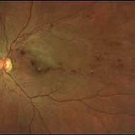
A zoomed vein occlusion in a young patient with systemic arterial hypertension.
Photographer: Gabriel Castilho, Suel Abujamra Institute, São Paulo.
Condition/keywords: branch retinal vein occlusion (BRVO), macular branch retinal vein occlusion (BRVO), non-perfused branch retinal vein occlusion (BRVO)
-
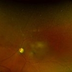
 Branches Starved of Flow, Yet Nature Strives to Grow
Apr 1 2025 by rohan jain
Tufts of NVE's in a case of Branch Retinal Vein Occlusion
Photographer: Dr. ROHAN JAIN
Condition/keywords: branch retinal vein occlusion (BRVO), capillary nonperfusion, non-perfused branch retinal vein occlusion (BRVO), non-perfusion, NVE, OCT Angiography, ST BRVO
-
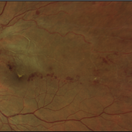
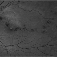
 Branch retinal vein occlusion - Colour & Red free image - ring shaped collaterals
Jul 18 2023 by Harsh Vardhan Singh, MS
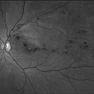
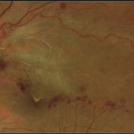
43-year-old woman presented with left eye old STBRVO with chronic CME of duration 6month showing ring shaped collaterals more evident on red free image
Photographer: Harsh Vardhan Singh, AIIMS, Guwahati
Imaging device: Zeiss Clarus 700
Condition/keywords: branch retinal vein occlusion (BRVO), BRVO, non-perfused branch retinal vein occlusion (BRVO)
-
 Branch retinal vein occlusion - Colour & Red free image - ring shaped collaterals
Jul 18 2023 by Harsh Vardhan Singh, MS
43-year-old woman presented with left eye old STBRVO with chronic CME of duration 6month showing ring shaped collaterals more evident on red free image
Photographer: Harsh Vardhan Singh, AIIMS, Guwahati
Imaging device: Zeiss Clarus 700
Condition/keywords: branch retinal vein occlusion (BRVO), BRVO, non-perfused branch retinal vein occlusion (BRVO)
-
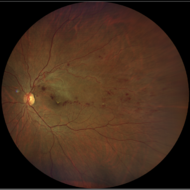
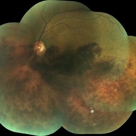 BRVO
BRVO
Apr 29 2018 by mahesh aryal
Fundus photo of 46-year-old man with BRVO montage photo.
Photographer: MAHESH ARYAL LUMBINI EYE INSTITUTE
Condition/keywords: non-perfused branch retinal vein occlusion (BRVO)
-
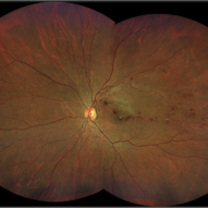
 BRVO - Color
Apr 10 2018 by Hosam Attia, MD
56-year-old African American male with ischemic nasal BRVO with advanced cupping OS - Optos California.
Imaging device: Optos California
Condition/keywords: branch vein occlusion (BVO), non-perfused branch retinal vein occlusion (BRVO)
-
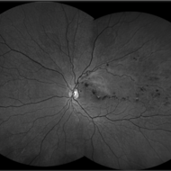
 BRVO - FA
Apr 10 2018 by Hosam Attia, MD
56-year-old African American male with ischemic nasal BRVO with advanced cupping OS - Optos California
Imaging device: Optos California
Condition/keywords: branch vein occlusion (BVO), non-perfused branch retinal vein occlusion (BRVO)
-
 BRVO With Non-Perfusion
May 3 2014 by Mallika Goyal, MD
Left eye superotemporal BRVO in a 53-year-old hypertensive male patient.
Photographer: Mallika Goyal, MD, Apollo Health City, Jubilee Hills, Hyderabad, India
Condition/keywords: non-perfused branch retinal vein occlusion (BRVO)
-
 BRVO With Non-Perfusion
May 3 2014 by Mallika Goyal, MD
Early phase fluorescein angiogram in an eye with superotemporal BRVO shows delayed filling of retinal vessels in the affected quadrant.
Photographer: Mallika Goyal, MD, Apollo Health City, Jubilee Hills, Hyderabad, India
Condition/keywords: non-perfused branch retinal vein occlusion (BRVO)
-
 BRVO With Non-Perfusion
May 3 2014 by Mallika Goyal, MD
Mid-phase fluorescein angiogram in an eye with superotemporal BRVO shows delayed filling of retinal vessels and non-perfusion in the affected quadrant.
Photographer: Mallika Goyal, MD, Apollo Health City, Jubilee Hills, Hyderabad, India
Condition/keywords: non-perfused branch retinal vein occlusion (BRVO)
-
 BRVO With Non-Perfusion
May 3 2014 by Mallika Goyal, MD
Fluorescein angiogram in an eye with superotemporal BRVO shows delayed filling of retinal vessels and non-perfusion in the affected quadrant.
Photographer: Mallika Goyal, MD, Apollo Health City, Jubilee Hills, Hyderabad, India
Condition/keywords: non-perfused branch retinal vein occlusion (BRVO)
-
 BRVO With Non-perfusion
May 3 2014 by Mallika Goyal, MD
Mid-phase fluorescein angiogram in an eye with superotemporal BRVO shows delayed filling of retinal vessels and non-perfusion in the affected quadrant.
Photographer: Mallika Goyal, MD, Apollo Health City, Jubilee Hills, Hyderabad, India
Condition/keywords: non-perfused branch retinal vein occlusion (BRVO)
-
 BRVO With Non-perfusion
May 3 2014 by Mallika Goyal, MD
Fluorescein angiogram in an eye with superotemporal BRVO shows delayed filling of retinal vessels and non-perfusion in the affected quadrant.
Photographer: Mallika Goyal, MD, Apollo Health City, Jubilee Hills, Hyderabad, India
Condition/keywords: non-perfused branch retinal vein occlusion (BRVO)
-
 BRVO With Non-perfusion
May 3 2014 by Mallika Goyal, MD
Late phase fluorescein angiogram in an eye with superotemporal BRVO shows delayed filling of retinal vessels and non-perfusion in the affected quadrant.
Photographer: Mallika Goyal, MD, Apollo Health City, Jubilee Hills, Hyderabad, India
Condition/keywords: non-perfused branch retinal vein occlusion (BRVO)
-
 BRVO With Non-perfusion
May 3 2014 by Mallika Goyal, MD
Late phase fluorescein angiogram in an eye with superotemporal BRVO shows delayed filling of retinal vessels and non-perfusion in the affected quadrant .
Photographer: Mallika Goyal, MD, Apollo Health City, Jubilee Hills, Hyderabad, India
Condition/keywords: non-perfused branch retinal vein occlusion (BRVO)
-
 BRVO With Non-perfusion
May 3 2014 by Mallika Goyal, MD
Late phase fluorescein angiogram of an eye with superotemporal BRVO shows delayed filling of retinal vessels, dilation and tortuosity of the affected veins, and non-perfusion in the affected quadrant .
Photographer: Mallika Goyal, MD, Apollo Health City, Jubilee Hills, Hyderabad, India
Condition/keywords: non-perfused branch retinal vein occlusion (BRVO)
 Loading…