Search results (103 results)
-
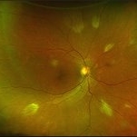
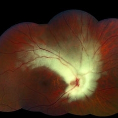
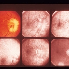
 Myelinated Nerve Fibers
Sep 17 2012 by Michael P. Kelly, FOPS
Retinal fundus photograph of myelinated nerve fibers
Photographer: Michael P. Kelly, FOPS Director, Duke Eye Labs, Duke University Hospital, Duke Eye Center
Imaging device: Topcon
Condition/keywords: myelinated nerve fibers
-
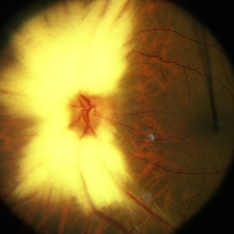
 Myelinated Nerve Fibre (MNF)
Jun 17 2023 by Harsh Vardhan Singh, MS
Fundus photograph of 32-year-old male having good best corrected visual acuity in both eyes with right eye having high myopia & MNF as incidental finding
Photographer: Dr Harsh Vardhan Singh, Assistant Professor, AIIMS, Guwahati
Condition/keywords: medullated nerve fibers, MNF, myelinated nerve fiber layer, myelinated nerve fibers, Nerve fiber layer arrangements, NFL
-
 Macular Hole With Myelinated Fibers L CF OD January 31, 2014
Mar 12 2014 by Manish Nagpal, MD, FRCS (UK), FASRS
Fundus photo of a 56-year-old woman having myelinated nerve fibers with a atrophic macular hole.
Photographer: Pooja Barot
Condition/keywords: macular hole, myelinated nerve fibers
-
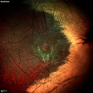
 Macular Pucker With Myelinated Nerve Fiber Layer
Nov 1 2018 by Kevin J. Blinder, MD, FASRS
Multi-color photo of macular pucker with myelinated nerve fiber layer.
Photographer: Jarrod Wehmeier
Imaging device: Heidelberg Spectralis
Condition/keywords: macular pucker
-
 Multiple Areas of Myelinated RNFL OD
Sep 18 2019 by John S. King, MD
68-year-old African American male presented with an acute PVD in the fellow eye. Fellow eye had similar findings, but the pics were not as good as OD.
Photographer: Brittany Dewberry
Imaging device: Optos CA
Condition/keywords: myelinated nerve fiber layer, myelinated nerve fibers
-
 Myelianated Nerve Fiber Layer
May 2 2013 by Henry J. Kaplan, MD
Extensive myelinated nerve fibers.
Condition/keywords: myelinated nerve fibers
-
 Myelinated Nerve Fibers
Apr 18 2025 by DR Rohit Gupta
The **myelinated nerve fibers of the optic disc** (also known as **medullated nerve fibers**) are retinal nerve fibers that retain their myelin sheath as they pass through the optic nerve head. Normally, retinal nerve fibers are unmyelinated to allow for light transparency, but in some cases, myelination extends anteriorly into the retina, appearing as a striking white, feathery patch on the optic disc or peripapillary retina. ### **Key Features:** 1. **Appearance:** - Dense, white, striated patches with feathery edges. - Typically located at the superior or inferior pole of the optic disc. - May obscure retinal vessels underneath. 2. **Clinical Significance:** - Usually **benign** and asymptomatic. - **Congenital** (present at birth or early childhood). - Rarely associated with **visual field defects** (e.g., scotomas corresponding to the area of myelination). - Occasionally linked with **high myopia** or **amblyopia** if extensive. 3. **Pathophysiology:** - Failure of oligodendrocytes or Schwann cells to stop myelination at the lamina cribrosa. - Normally, myelination stops at the optic nerve head, but in this condition, it extends into the retina. 4. **Diagnosis:** - **Fundoscopy:** Classic white, feathery appearance. - **Optical Coherence Tomography (OCT):** Shows thickened retinal nerve fiber layer (RNFL). - **Visual Field Testing:** May detect defects if large. 5. **Differential Diagnosis:** - Optic disc edema - Cotton wool spots - Retinoblastoma (rarely, but must be ruled out in children) 6. **Management:** - No treatment required if asymptomatic. - Monitor for amblyopia in children. - Rare cases with significant visual impairment may need further evaluation. ### **Fun Fact:** Myelinated nerve fibers are seen in **~0.5-1%** of the population and are usually an incidental finding.
Photographer: Dr Rohit gupta
Imaging device: Samsung S21
Condition/keywords: Medulated Nerve fibre, Medullated Nerve fibres, myelinated nerve fibers, Myelinated Nerve Fibres, optic disc drusen
-
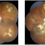 NPDR With Myelinated Nerve Fibers
Nov 5 2018 by Diva Kant Misra, MBBS, DO, DNB, MNAMS, FVRS
Bilateral montage funds photo images of a 56-year-old diabetic patient showing signs of NPDR along with myelinated nerve fibers.
Photographer: Hiteshwar Saikia
Condition/keywords: diabetes, hard exudates, myelinated nerve fibers, nonproliferative diabetic retinopathy
-
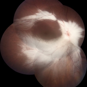
 Unilateral Myelinated Nerve Fiber
Nov 12 2019 by Aditya S Kelkar, MS, FRCS, FASRS,FRCOphth
Left eye fundus photograph of a 29-year-old male with extensive myelinated retinal nerve fibers appearing as grey white opaque lesions on the retina with feathery edges obscuring the retina.
Photographer: Dr. Vikrant Narwade
Condition/keywords: myelinated nerve fibers
-
---thumb.JPG/image-square;max$300,300.ImageHandler) Myelinated Nerve Fiber Layer - Fundus Image
Myelinated Nerve Fiber Layer - Fundus Image
Oct 5 2013 by Roy Schwartz, MD
Left eye of a 20-year-old female with myelinated nerve fibers.
Photographer: Galit Yair-Pur
Condition/keywords: fundus photograph, myelinated nerve fibers
-
 Myelinated Nerve Fiber With Epiretinal Membrane With Lamellar Macular Hole
May 4 2020 by SWATI INDURKHYA
Heidelberg HRA + OCT Spectralis Multicolor (30 degree) retinal image of the left eye of a 28-year-old male showing myelinated nerve fibre (MNF) with epiretinal membrane (ERM) and lamellar macular hole (LMH).
Photographer: Rakesh PR, Giridhar Eye Institute, Kerala, India
Imaging device: Heidelberg Spectralis HRA + OCT
Condition/keywords: epiretinal membrane (ERM), myelinated nerve fibers
-
 Choroidal Osteoma Associated to Myelinated Nerve Fibers
Sep 11 2019 by Sophia El Hamichi, MD
A 36-year-old male with supratemporal juxta papillary choroidal osteoma associated with myelinated nerve fibers of the left eye.
Photographer: Sophia El Hamichi, MD, Murray Ocular Oncology and Retina
Imaging device: Optos
Condition/keywords: choroidal osteoma, myelinated nerve fibers, Optos
-
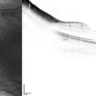 Myelinated Nerve Fiber Layer - OCT Image
Oct 5 2013 by Roy Schwartz, MD
Left eye of a 20-year-old female with myelinated nerve fibers
Photographer: Galit Yair-Pur
Condition/keywords: myelinated nerve fibers, optical coherence tomography (OCT)
-
 Myelinated Nerve Fibers
Sep 17 2012 by Michael P. Kelly, FOPS
Retinal fundus photograph of a macular hole.
Photographer: Michael P. Kelly, FOPS Director, Duke Eye Labs, Duke University Hospital, Duke Eye Center
Imaging device: Topcon
Condition/keywords: macular hole, myelinated nerve fibers
-
 Myelinated Nerve Fibers
Feb 12 2015 by Timothy S Fuller, MD
Fundus photograph of a 20-year-old woman with myelinated nerve fibers.
Photographer: Nick Hesse, Texas Retina Associates
Condition/keywords: myelinated nerve fibers
-
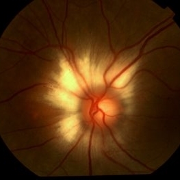
 Myelinated nerve fibres
Jan 11 2013 by Alex P. Hunyor, MD
Myelinated nerve fibres at optic disc, left eye.
Condition/keywords: myelinated nerve fibers
-
 Myelinated Retinal Nerve Fiber Layer
Jan 26 2020 by Prithvi Chandrakanth
Incidental fundus finding in an asymptomatic 37-year-old female of grayish white , well demarcated patch, with frayed borders obscuring underlying vessels at the optic nerve head in both eyes.
Photographer: Dr.PRITHVI CHANDRAKANTH, ARAVIND EYE HOSPITAL, UDUMALPET
Imaging device: TRASH TO TREASURE RETCAM
Condition/keywords: myelinated nerve fibers, retcam, smartphone fundus photography
-
 Myelinated Nerve Fiber Layer
Apr 3 2018 by Mitzy E Torres Soriano, MD
Myelinated nerve fiber layer, left eye.
Photographer: Mitzy Torres Soriano
Condition/keywords: myelinated nerve fiber layer, myelinated nerve fibers
-
 Myelinated retinal nerve fibers Slide 2
Oct 22 2012 by Ronald C. Gentile, MD
A magnified fundus photo of the myelinated retinal nerve fibers showing the retinal vascular abnormalities with multiple aneurysmal dilations.
Photographer: The New York Eye & Ear Infirmary Department of Medical Imaging
Condition/keywords: myelinated nerve fibers, retinal telangiectasia
-
 AO
AO
Sep 3 2013 by Howard Schatz, MD
Myelinated nerve fibers, AO, BAO.
Condition/keywords: AO, myelinated nerve fibers
-
---thumb.jpg/image-square;max$300,300.ImageHandler) ARMD RPE Defect / Myelinated NFL
ARMD RPE Defect / Myelinated NFL
Jan 9 2014 by David Callanan, MD
ARMD RPE defect , myelinated nerve fiber layer in a 47-year-old male patient.
Condition/keywords: myelinated nerve fiber layer, retinal pigment epithelium (RPE) defect
 Loading…