Search results (561 results)
-
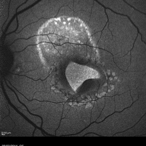
 Retinal Detachment with PVR (s/ SPR, PPV, MPV, 360 Retinectomy, PFO, PI, FAx, SO)
Aug 22 2019 by Merrick Avila
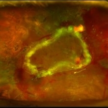
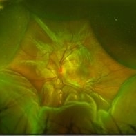
Ultra-wide field pseudocolor fundus photograph of a 64-year-old female with a treated retinal detachment with proliferative vitreoretinopathy. Patient has a history of complex retinal detachments that have been treated multiple times. On exam 8-22-19, there were large macular holes with LP vision. There was a long discussion about guarded nature of her condition and goals or trial for repair including globe sparing prevention of phthisis.
Photographer: Merrick Avila
Imaging device: Optos
Condition/keywords: diabetic retinopathy, hemorrhage, Optos, proliferative vitreoretinopathy (PVR), retinectomy, silicone oil
-
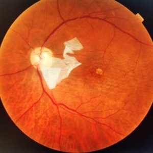
 Green Goblin Detachment
Jan 13 2022 by Netan Choudhry, MD, FRCS(C) FASRS
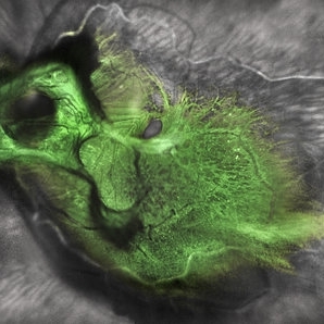
Tractional retinal detachment with macular hole in a 76-year-old female.
Photographer: John Golding BA, Vitreous Retina Macula Specialists of Toronto, OCTane Imaging Lab
Imaging device: Multicolor fundus photo taken on the Spectralis OCT2 (Heidelberg Engineering GmbH).
Condition/keywords: macular hole, Multispectral imaging, tractional retinal detachment
-
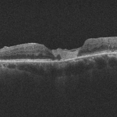
 Myopic Traction Maculopathy
Mar 17 2025 by Drew Mitchell
HD 1 line 100x 9 mm scan of a right eye with MTM at stage 3c. Macular Schisis Detachment.
Photographer: Drew Mitchell OCT-C
Imaging device: Zeiss Cirrus 5000
Condition/keywords: full thickness macular hole, Macular hole, myopic foveoschisis, myopic macular schisis, myopic traction maculopathy, PVD
-
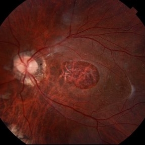
 Retinitis Pigmentosa with Macular Hole with Posterior Subcapsular Cataract
Apr 28 2025 by Malvika Singh
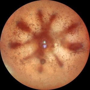
Fundus photograph of the right eye of a 31 year old with retinitis pigmentosa with a macular hole, showing the shadow of posterior subcapsular cataract over the fundus.
Photographer: Dr Malvika Singh, Retina Foundation, Ahmedabad, India
Imaging device: Mirante SLO/OCT
Condition/keywords: macular hole, posterior subcapsular cataract, retinitis pigmentosa
-
 Intravitreal Cysticercosis With Full Thickness Macular Hole
Apr 30 2018 by Vishal Agrawal, MD, FRCS,FACS,FASRS
Fundus montage picture of a 40-year-old man presenting with decreased vision in the right eye for the past 2 months. Live intravitreal cysticercosis can be seen lying on the retina. Zooming the image reveals the full thickness macular hole. The scolex invaginates with the light of the camera causing double image of the cyst because of movement .
Photographer: Vishal Agrawal MD,FRCS
Imaging device: Zeiss 524
Condition/keywords: cysticercosis, full thickness macular hole
-
 Macular Tear
May 14 2014 by Avris Romario Diparaja Siahaan
Blue autofluorescence (BAF) a 40-year-old man with macular tear (had a photocoagulation laser).
Photographer: Avris Romario Diparaja Siahaan
Imaging device: Heidelberg HRA + OCT Spectralis
Condition/keywords: autofluorescence imaging, macular hole
-
 Tractional Detachment of Retina
Aug 21 2024 by Jordyn Beckman
18 year old male with tractional detachment of Retina, chronic macular hole and silicone oil s/p RD repair x2. BCVA CF @2 ft, fellow eye prosthetic.
Photographer: Jordyn Beckman
Imaging device: Optos California
Condition/keywords: Macular hole, preretinal fibrosis, Retinal Detachment, scleral buckle, silicone oil, TRACTION, tractional retinal detachment
-
 AMG on Papillomacular Bundle
Mar 16 2025 by PUJA NEGI
Patient came to our OPD with history of AMG done for macular hole . On examination we found that the AMG had displaced over the papillomacular bundle from the hole.
Photographer: DR Nuzhat
Condition/keywords: amniotic membrane graft, macular hole
-
 Amniotic-Membrane Grafted Macular Hole
Oct 25 2023 by Jessica Hampton, BS
Optical-coherence tomography image of a 67-year old woman with a recurrent, chronic full-thickness macular hole in the left eye repaired with an amniotic membrane graft, seen at 2 years follow up.
Photographer: Dr. Diana Do, Stanford Medicine, Byers Eye Institute
Condition/keywords: amniotic membrane graft, full thickness macular hole
-
 Choroidal and Retinal Detachment Secondary to Full-thickness Macular Hole
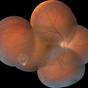
May 24 2024 by Tony Y Chen, MD
Optos photograph of a 61-year-old woman with choroidal and retinal detachment secondary to full-thickness macular hole.
Condition/keywords: choroidal detachment, Retinal detachment with macular hole
-
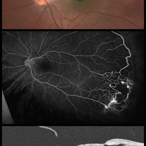 Coats Disease - Fundus Image, Angiography and OCT
Feb 3 2021 by Gabriel Costa Andrade, PhD
Fundus image, angiography and OCT of a 12-year-old boy with coats disease, pheripheral neovascularization and macular hole.
Photographer: Dr Gabriel Andrade, Dr André Maia
Condition/keywords: Coats' disease
-
 Full Thickness Macular Hole With ERM
Feb 26 2014 by Sharon Fekrat, MD FACS FASRS
Middle aged woman with a full thickness macular hole in the left eye associated with an epiretinal membrane.
Photographer: Michael P Kelly, Ophthalmic Photographer, Duke Eye Imaging, Duke Eye Center
Condition/keywords: epiretinal membrane (ERM), macular hole
-
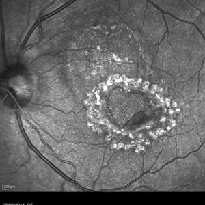
 Giant Persistent Macular Hole
Dec 6 2024 by César Adrián Gómez Valdivia, MD
Giant Persistent Macular Hole found in a 48 year-old male patient one year after vitrectomy.
Photographer: @eyemissu2
Imaging device: TOPCON TRC-50DX
Condition/keywords: macular, macular hole
-
Iatrogenic Macular Hole and Subretinal Migration of PFCL
Feb 7 2023 by Aditya S Kelkar, MS, FRCS, FASRS,FRCOphth
The video demonstrates a surgical scenario where the fovea gives away by the force imparted by the jet of an injecting PFCL (Perfluorocarbon heavy Liquid) and the PFCL migrates subfoveally. Intraoperative OCT confirms the presence of a macular hole. The situation is managed by ILM peeling and mobilizing subfoveal PFCL peripherally by injecting another bubble of PFCL over the posterior pole. A peripheral drainage retinotomy is then created to aspirate the subretinal PFCL followed by fluid-air exchange, PFCL-air exchange, and endolaser around the retinotomy. Post-operative OCT at 3 weeks’ follow-up shows a sealed macular hole.
Condition/keywords: Iatrogenic macular hole, Intraoperative complications, Subretinal PFCL
-
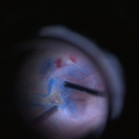
 ILM Peeling in Progress
Feb 4 2022 by Manish Nagpal, MD, FRCS (UK), FASRS
Intraoperative shot of ILM peeling in progress using forceps.
Photographer: Manish Nagpal, Director, Retina Foundation, Ahmedabad
Imaging device: Sony PMW -10 MD surgical camera
Condition/keywords: ILM flap, ILM staining, internal limiting membrane (ILM) peeling, macular hole, retina, retina surgery
-
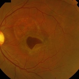
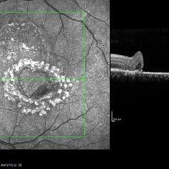
 Macular Hole
Mar 29 2013 by Henry J. Kaplan, MD
Chronic macular hole with drusen like deposits and surrounding cuffing of subretinal fluid.
Condition/keywords: macular hole
-
 Macular Hole Due to Proliferative Diabetic Retinopathy
Aug 13 2025 by Ricardo Leitão Guerra
A macular hole formation after anti-VEGF injection prior to vitrectomy for tractional retinal detachment in a patient presenting proliferative diabetic retinopathy.
Photographer: Ricardo Leitão Guerra
Imaging device: ZEISS CLARUS 700
Condition/keywords: macular hole, proliferative diabetic retinopathy (PDR)
-
Macular Hole Repair Under PFCL
Jun 6 2024 by DAVID PÉREZ GONZÁLEZ, MD
The heavy liquid, though slightly more expensive, can be a valuable tool for macular hole repair. When applied to the posterior area, it can stabilize the hole's edges, facilitating its position and draining the adjacent subretinal fluid.
Condition/keywords: ILM peeling, macular hole, Perfluorocarbon Liquid, PFCL, vitrectomy
-
 Macular Hole With Folded ILM
Mar 12 2016 by Sjakon G Tahija, MD
This is a freeze frame taken from a large vitrectomy for macular hole where I folded the ILM in a cone around the hole before fluid air exchange.
Photographer: Sjakon Tahija
Imaging device: Sony
Condition/keywords: macular hole, pars plana vitrectomy (PPV)
-
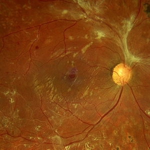
 Macular Hole With Myelinated Fibers L CF OD January 31, 2014
Mar 12 2014 by Manish Nagpal, MD, FRCS (UK), FASRS
Fundus photo of a 56-year-old woman having myelinated nerve fibers with a atrophic macular hole.
Photographer: Pooja Barot
Condition/keywords: macular hole, myelinated nerve fibers
-
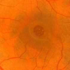
 Macular Pseudohole
Feb 7 2017 by Manish Nagpal, MD, FRCS (UK), FASRS
This OCT reveals the central break in the hyaloid which clinically mimics a macular hole.
Photographer: pooja barot
Condition/keywords: macular pseudohole
-
 Macular Pseudohole
Feb 7 2017 by Manish Nagpal, MD, FRCS (UK), FASRS
Fundus appearance of a macular hole is basically a small central break in the hyaloid mimicking a macular hole. The OCT in the next picture depicts the break in the hyaloid.
Photographer: Pooja Barot
Condition/keywords: macular pseudohole
-
 Macular Tear
May 14 2014 by Avris Romario Diparaja Siahaan
Fundus photograph a 40-year-old man with macular tear (had a photocoagulation laser).
Photographer: Avris Romario Diparaja Siahaan
Imaging device: Topcon TRC 50 DX Type IA
Condition/keywords: macular hole
-
 Macular Tear
May 14 2014 by Avris Romario Diparaja Siahaan
Infrared photograph a 40-year-old man with macular tear (had a photocoagulation laser).
Photographer: Avris Romario Diparaja Siahaan
Imaging device: Heidelberg HRA + OCT Spectralis
Condition/keywords: infrared image, macular hole
-
 Macular Tear
May 14 2014 by Avris Romario Diparaja Siahaan
OCT a 40-year-old man with macular tear (had a photocoagulation laser).
Photographer: Avris Romario Diparaja Siahaan
Imaging device: Heidelberg HRA + OCT Spectralis
Condition/keywords: macular hole, optical coherence tomography (OCT)
 Loading…