Search results (18 results)
-
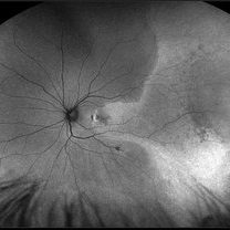
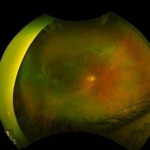
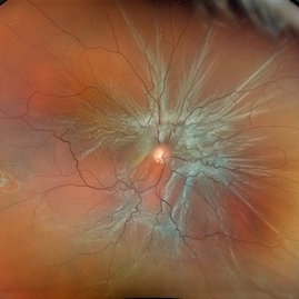
 Macula Off Retinal Detachment
Mar 22 2023 by Zach Seim
An ultra-widefield fundus image of a 65 year old male with a Macula Off Retinal Detachment. Patient's vision at the time of the image was CF at 6 Feet and surgical options were discussed. Fluid-gas exchange was performed without complications.
Photographer: Zach Seim
Imaging device: Optos California
Condition/keywords: left eye, macula off retinal detachment, OPTOS CALIFORNIA, scanning laser ophthalmoscope, ultra-widefield image
-
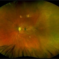
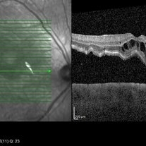 Macula Off Retinal Detachment
Jan 2 2018 by Carolyn Daley
55-year-old with macula off retinal detachment post cataract surgery.
Photographer: Carolyn Daley, Retina Specialists of Michigan
Imaging device: Heidelberg Spectralis
Condition/keywords: Heidelburg Spectralis, optical coherence tomography (OCT)
-
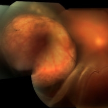
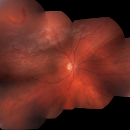
 Total retinal Detachment multiple holes
Sep 26 2022 by Denica Rodriguez
60 year old Male presented with two week old Macula off Retinal detachment with multiple tears.
Photographer: Denica Rodriguez
Imaging device: Optos California
Condition/keywords: color fundus photograph, color photo, macula-off, optos, pseudocolor, Retinal detachment, retinal holes, retinal tear, Retinal tear with detachment, superior arcade, superior field, superior retina, total retinal detachment
-
 Choroidal Melanoma With a Serous Retinal Detachment
Aug 23 2018 by Nichole Lewis
63-year-old male with a large choroidal melanoma and a serous macula off retinal detachment. Vision is count fingers.
Photographer: Nichole Lewis
Condition/keywords: serous retinal detachment
-
 Macula off Retinal Detachment
Jan 23 2024 by Annaka Gooding
Ultra-widefield fundus photograph of an 81-year-old male with a Macula Off Retinal Detachment affecting his right eye. Patient presented at office with complaints of flashes of light for about 2 weeks accompanied by a curtain veil covering inferior visual field. Patient had total vision loss 24 hours prior to visit. His vision was scHM. The physician recommended Retinal Detachment Repair with PPV.
Photographer: Annaka Gooding, CPO
Imaging device: Optos California
Condition/keywords: detachment, fundus photography, macula off retinal detachment, Optos, retinal detachment of the macula, right eye, ultra-wide field imaging
-
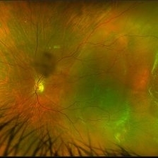 Macula Off Retinal Detachment
Jun 23 2023 by Zach Seim
Ultra-widefield fundus photograph of a 42 year old women with a Macula Off Retinal Detachment in her left eye. At the time of this photograph the patient's visual acuity was sc CF at 2 feet.
Photographer: Zach Seim
Imaging device: Optos California
Condition/keywords: left eye, macula off retinal detachment, Optos, OPTOS CALIFORNIA, scanning laser ophthalmoscope, ultra-widefield image
-
 Macula Off Retinal Detachment
Jun 25 2024 by Zach Seim
Optos Fundus photo of a 47 year old female with a Macula Off Retinal Detachment right eye, presenting with loss of nasal visual field. Patient's vision at presentation was DCC 20/100-1. Patient was counseled and decided to proceed with surgery.
Photographer: Zach Seim
Imaging device: OPTOS California
Condition/keywords: macula off retinal detachment, Optos, OPTOS CALIFORNIA, right eye
-
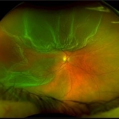
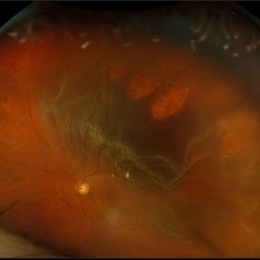
 Macula Off Retinal Detachment with CNV
Nov 11 2019 by Olivia Rainey
Ultra-wide field pseudocolor photograph of a 42-year-old female with a long-standing, macula-off retinal detachment affecting her left eye. Patient was unaware of vision loss until testing her visual acuity and she denied seeing flashing lights. Patient decided to proceed with scleral buckling. The CNV is potentially secondary the retinal detachment, but may be myopic related or idiopathic. The CNV appears fibrotic and inactive. The patient was warned that this will absolutely limit how much vision she recovers once the retina is reattached.
Photographer: Olivia Rainey
Imaging device: Optos California
Condition/keywords: choroidal neovascularization (CNV), chronic retinal detachment, fundus autofluorescence (FAF), left eye, montage, Optos, retinal detachment of the macula, ultra-wide field imaging
-
 Macula Off Retinal Detachment with CNV
Nov 11 2019 by Olivia Rainey
Ultra-wide field pseudocolor photograph of a 42-year-old female with a long-standing, macula-off retinal detachment affecting her left eye. Patient was unaware of vision loss until testing her visual acuity and she denied seeing flashing lights. Patient decided to proceed with scleral buckling. The CNV is potentially secondary the retinal detachment, but may be myopic related or idiopathic. The CNV appears fibrotic and inactive. The patient was warned that this will absolutely limit how much vision she recovers once the retina is reattached.
Photographer: Olivia Rainey
Imaging device: Optos California
Condition/keywords: choroidal neovascularization (CNV), left eye, montage, Optos, pseudocolor, retinal detachment of the macula, ultra-wide field imaging
-
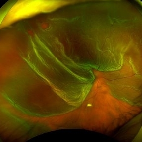
 Macula Off Retinal Detachment with Horseshoe Tear
May 10 2017 by Nichole Lewis
Macula off retinal detachment with horseshoe tear.
Photographer: Nichole Lewis
-
 OD-Macula Off Retinal Detachment
Sep 9 2020 by Lauren Schuler
Optos photo of a 71-year-old male with a macula (mac off) off retinal detachment of the right eye.
Photographer: Lauren Schuler
Imaging device: Optos
Condition/keywords: detachment, macula, retina
-
 Peripheral Retinoschisis
Feb 12 2020 by DIEGO TOLENTINO
Peripheral retinoschisis plus macula off retinal detachment.
Photographer: Diego Tolentino, CEOP
Condition/keywords: retinal detachment of the macula, senile retinoschisis
-
 Retinal Detachment Preop and Postop Day-1
Feb 6 2025 by Sham Talati, DOMS
Sharing the pre-operative and the post-operative day-1 fundus photos of a case of a 20 year old high myopic female who presented to our hospital with C/O loss of vision in her Right Eye. On examination she was diagnosed with Retinal Detachment in her Right eye. She was operated on same day with Scleral Buckle surgery. After the successful scleral buckle surgery the very next day the retina is well attached and patient got back her lost vision.
Photographer: Dr. Sham Talati , Dr. Talati's Eye Hospital , Ahmedabad
Condition/keywords: macula off retinal detachment, myopic retinal retachment, Retinal Detachment, scleral buckle
-
 Retinal Detachment with Proliferative Vitreoretinopathy
Jan 31 2022 by Ahmad B. Tarabishy, MD
Ultra wide-field fundus photograph of a 55-year-old gentleman who had previously underwent laser retinopexy for multiple inferior retinal breaks. He presented with a macula-off retinal detachment from a new temporal break with proliferative vitreoretinopathy with fixed folds noted temporally and superonasally.
Photographer: Megan McLandsborough, Lakeland Eye Clinic
Imaging device: Optos California UWF Camera
Condition/keywords: laser retinopexy, macula off Retinal Detachment, proliferative retinopathy, proliferative vitreoretinopathy (PVR), Retinal Detachment, retinal detachment with retinal defect
-
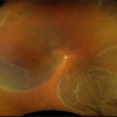
 Rhegmatogenous Macula Off Retinal Detachment with Multiple Breaks
May 29 2024 by Alexis Singstock
Ultra widefield fundus photograph of a 66 year old male with rhegmatogenous macula off retinal detachment with multiple breaks. Patient presented emergently for a curtain/veil in inferonasal visual field. Patient reports the curtain/veil in left eye started about 1 week prior, yet denied seeing flashes and floaters. Patient's vision was hand motion. Dr. Edward Korot examined the patient and scheduled him for a scleral buckle along with pars plana vitrectomy surgery.
Photographer: Alexis Singstock, Retina Specialists of Michigan
Imaging device: Optos California
Condition/keywords: fundus photography, left eye, macula off retinal detachment, OPTOS CALIFORNIA, pars plana vitrectomy (PPV), rhegmatogenous retinal detachment, scleral buckle, ULTRA WIDE FIELD
-
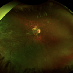
 Rhegmatogenous Macula-Off Retinal Detachment
Sep 19 2024 by Alexis Singstock
Ultra wide field fundus photograph of a 62 year old female with a rhegmatogenous macula-off retinal detachment affecting her right eye. Patient reported decreased vision, curtain/veil in vision and eye pain with the onset approximately 2 weeks prior to initial encounter.
Photographer: Alexis Singstock
Imaging device: Optos California RGB
Condition/keywords: macula off retinal detachment, Optos, OPTOS CALIFORNIA, OPTOS CALIFORNIA RGB, Retina detachment, ultra-wide field imaging
-
 ROP related retinal detachment
Jun 8 2022 by Alexandre Grandinetti, MD, PhD
16-year-old girl with macula off retinal detachment and ROP cicatricial changes. She was born with 28 weeks of gestation and had no treatment
Photographer: Corina Skrzek, Hospital de Olhos do Paraná
Imaging device: California
Condition/keywords: rop retinal detachment
-
 Twinkle Twinkle
Aug 5 2024 by Virginia Gebhart
65 year old male with mac-off retinal detachment with 360 folds and horseshoe tear.
Photographer: Virginia Gebhart
Imaging device: Optos California
Condition/keywords: macula off retinal detachment, RD, Retinal Detachment
 Loading…