Search results (31 results)
-
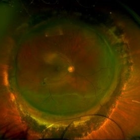
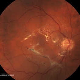
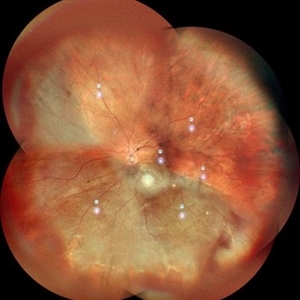
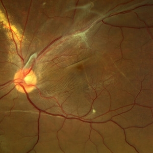 Displaced & folded macula
Oct 10 2022 by Ricardo Leitão Guerra
Tractional retinal detachment due to sickle cell retinopathy leading to a displaced and folded appearance of the macula in this 36-yo male. Subretinal bands are also noticed crossing the macula towards inferior retinal detachment area.
Photographer: Ricardo Leitão Guerra
Imaging device: Clarus 700 - Zeiss
Condition/keywords: folds, sickle cell retinopathy, subretinal bands, tractional retinal detachment
-
 Dexamethasone Implant
Jul 3 2021 by Gerardo Rivera Arroyo
42-year-old male, operated on for vitrectomy plus scleral buckling plus silicone plus dexamethasone implant for inferior retinal detachment with PVR.
Photographer: Rosa Elizabeth Moreno Anda, MD, Hospital Central Militar CDMX
Condition/keywords: dexamethasone implant, retina surgery, vitrectomy
-
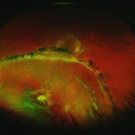
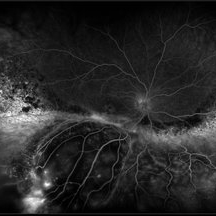 Exudative Retinal Detachment and Branch Retinal Vein Occulsion
Oct 29 2020 by Olivia Rainey
Ultra-widefield fluorescein anigogram of a 51-year-old female with an exudative retinal detachment and branch retinal vein occlusion with retinal neovascularization affecting her right eye. The physician stated that the multiple aneurysmal dilations noted in the inferior periphery are responsible for the exudative RD seen on exam. He is considering Coat's vs FEVR given family history of aneurysms/congenital heart pathology per patient. He encouraged the patient to control their blood pressure, cholesterol, blood sugar, and co-morbidities which may have promoted the BRVO. He recommended antiVEGF injections to control the vascular leakage. Given the severe presentation and imminent threat to her vision, he recommended Eylea as first line therapy.
Photographer: Olivia Rainey, OCT-C, COA
Imaging device: Optos California
Condition/keywords: branch retinal vein occlusion (BRVO), chronic retinal detachment, fluorescein angiogram (FA), fluorescein leakage, inferior retina, inferior retinal detachment, Optos, ultra-wide field imaging
-
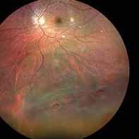 Inferior retinal detachment with lattice and holes
May 31 2023 by Aditya S Kelkar, MS, FRCS, FASRS,FRCOphth
Importance of dilated retina check up before Lasik surgery can't be better demonstrated...patient totally asymptomatic came for Lasik opinion and has inferior retinal detachment with lattice and holes, sparing the macula
Photographer: Dr. Sahil Wagh , National Institute of Opthalmology, Pune , India
Imaging device: Zeiss Clarus 500
Condition/keywords: inferior retinal detachment
-
 Retinal detachment
Apr 12 2023 by Ahmed Abbas Hashmi, OD
Color fundus photograph of the left eye of a 30-year-old man with asymptomatic inferior retinal detachment with pigmented demarcation line. Macula and Disc healthy.
Photographer: Ahmed Abbas Hashmi
Imaging device: Topcon TRC-NW8F
Condition/keywords: Pigmentary demarcation line, Retinal Detachment
-
 Retinal Detachment After Retinoblastoma Treatment
Mar 10 2024 by Alexandre Grandinetti, MD, PhD
Inferior retinal detachment occurring 6 years after treatment with intraarterial chemotherapy and laser in an 8-year-old boy.
Photographer: Corina Szrek
Condition/keywords: pediatric, retinoblastoma
-
 Retinal Detachment Sparing Fovea By Microns
Sep 24 2018 by samarth mishra
A 29-year-old young female presented with complaint of blurring of vision in the right eye since one year. Best corrected visual acuity was 20/40. On routine examination inferior retinal detachment was noted. Optical coherence tomography (OCT) showed the retinal detachment sparing the fovea by few microns.
Photographer: Aditya Birla Sankara Nethralaya, Kolkata , West Bengal , India
Condition/keywords: color fundus photograph, multicolor, optical coherence tomography (OCT)
-
 Scleral Buckling IOL Drop
Aug 6 2023 by Dr.Sheetal Divate
A 27 year old female with an old history of trauma and operated with scleral buckling and cataract surgery in the past came recently with complaints of DOV . Findings noted where IOL drop, inferior retinal detachment and old scleral buckle indent.
Photographer: Dr.Sheetal Divate
Imaging device: Optos Advance
Condition/keywords: dislocated intraocular lens (IOL), Retinal Detachment, scleral buckle
-
 Chronic Inferior Retinal Detachment
Mar 1 2017 by Philip J. Polkinghorne, MD
Color photograph of chronic retinal detachment with pigment demarcation line and atrophic holes visible. The vision was recorded at 20/20, and follow up is 3 years.
Photographer: Alex Fraser
Condition/keywords: atrophic retinal hole, demarcation line
-
 Old RRD With Retinal Cysts and High Watermark
Apr 10 2020 by Dipak Nag, MBBS, FCPS, MSc, FRF
Intra-operative fundus picture of a 20-year-old boy showing multiple retinal cysts and high watermark in a case of old inferior retinal detachment OD.
Photographer: Dipak
Condition/keywords: high watermark, retinal cyst
-
 Inferior retinal detachment
Dec 19 2012 by Eric A. Postel, MD
Color fundus photograph of an inferior retinal detachment
-
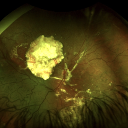
 Choroidal Mass
Mar 4 2024 by ANKIT JAIN
Left eye color photo montage of 39 year old female with sub retinal mass in nasal quadrant with hemorrhages and subretinal fluid with inferior retinal detachment.
Photographer: Dr Ankit Jain
Imaging device: MIRANTE
Condition/keywords: choroidal mass
-
 Chronic Inferior Retinal Detachment
Mar 1 2017 by Philip J. Polkinghorne, MD
Fundus autofluorescence of a chronic inferior retinal detachment.
Photographer: Alex Fraser
Condition/keywords: fundus autofluorescence (FAF)
-
 Chronic Retinal Detachment: Features Slide 1
Oct 22 2012 by Ronald C. Gentile, MD
Chronic retinal detachments can be associated with demarcation lines (tidemarks), subretinal bands or sheets, and retinal cysts. Fundus photo of a chronic inferior retinal detachment reveals multiple demarcation lines inferior to the center of the fovea as a result of an inferior temporal dialysis.
Photographer: The New York Eye & Ear Infirmary Department of Medical Imaging
Condition/keywords: chronic retinal detachment, demarcation line
-
 Inferior RD OD with Exposed Scleral Buckle OD in Previous Images
Feb 4 2013 by James B. Soque, CRA, OCT-C, COA, FOPS
Fundus image of 66-year-old WM with Hx of SBOD in 2009. presents with exposed SBOD and infection seen in accompanying images.
Photographer: James Soque, CRA COA
Imaging device: Topcon TRC 50 DX, MERGE Imaging Software
Condition/keywords: inferior retinal detachment
-
 Inferior Retinal Detachment with Lattice
Sep 30 2020 by Sham Talati, DOMS
A patient of inferior retinal detachment with lattice inferiorly.
Photographer: Dr. Sham Talati,Retina Foundation,Ahmedabad
Imaging device: Nidek Mirante
Condition/keywords: lattice degeneration
-
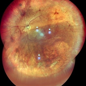
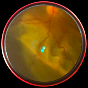
 Juvenile Retinoschisis with Localized Inferior Retinal Detachment
May 4 2020 by Giridhar Anantharaman, MS
Optos ultra-widefield retinal image of the left eye of a 11-year-old male child showing peripheral retinoschisis with localized inferior retinal detachment.
Photographer: Rakesh PR , Giridhar Eye Institute, Kerala, India
Imaging device: Optos UWF Daytona Plus
Condition/keywords: retinoschisis
-
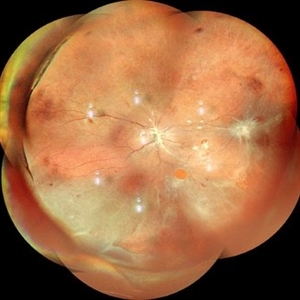
 Pars Planitis - Peripheral Uveitis
Nov 9 2012 by Norman Byer
This 25-year-old man had pars planitis, peripheral uveitis bilaterally. In this eye it produced a small tractional oval tear of the retina and an inferior retinal detachment. The typical creamy yellow exudates of pars planitis can be seen in the lower right very close to the ora serrata.
Condition/keywords: creamy yellow exudates, inferior retinal detachment, pars planitis, peripheral uveitis, tractional retinal tear
-
 Retinal Detachment
May 13 2016 by Nichole Lewis
Inferior Retinal Detachment with some demarcation line s/p barrier laser.
Photographer: Nichole Lewis
Condition/keywords: barrier laser
-
 Retinal Detachment
Apr 28 2024 by Anjana Mirajkar, MS Ophthalmology
A montage of a 40 year old male showing multiple breaks with inferior retinal detachment with peripheral traction in a silicon filled eye.
Photographer: Dr. Anjana Mirajkar -Retina Foundation, Ahmedabad
Imaging device: Mirante-Nidek
Condition/keywords: Retinal detachment under Silicon Oil
-
 Retinal Detachment
Nov 3 2023 by Anjana Mirajkar, MS Ophthalmology
A widefield image of OS of a 55 year old female case of inferior retinal detachment with macula off.
Photographer: Dr. Anjana Mirajkar -Retina Foundation, Ahmedabad
Imaging device: Mirante-Nidek
Condition/keywords: inferior retinal detachment, Retinal Detachment
-
 Retinal Detachment
Nov 3 2023 by Anjana Mirajkar, MS Ophthalmology
A widefield image (montage) of OS of a 55 year old female case of inferior retinal detachment with macula off.
Photographer: Dr. Anjana Mirajkar -Retina Foundation, Ahmedabad
Imaging device: Mirante-Nidek
Condition/keywords: inferior retinal detachment, Retinal Detachment
-
 Retinal Detachment - Smartphone Fundus Photography
Jun 10 2019 by Prithvi Chandrakanth
Asymptomatic 65-year-old female, an incidental finding of inferior retinal detachment on evaluation for cataract surgery.
Photographer: Dr.Prithvi Chandrakanth, Dr.Chandrakanth Malabar Nethralaya, Kozhikode, India.
Imaging device: TRASH TO TREASURE RETCAM
Condition/keywords: retina, smartphone fundus photography
-
 Retinal Detachment Sparing Fovea By Microns
Sep 24 2018 by samarth mishra
A 29-year-old young female presented with complaint of blurring of vision in the right eye since one year. Best corrected visual acuity was 20/40. On routine examination inferior retinal detachment was noted. Optical coherence tomography (OCT) showed the retinal detachment sparing the fovea by few microns.
Photographer: Aditya Birla Sankara Nethralaya, Kolkata , West Bengal , India
Condition/keywords: color fundus photograph, multicolor, optical coherence tomography (OCT)
-
 Retinal Detachment Sparing Fovea By Microns
Sep 24 2018 by samarth mishra
A 29-year-old young female presented with complaint of blurring of vision in the right eye since one year. Best corrected visual acuity was 20/40. On routine examination inferior retinal detachment was noted. Optical coherence tomography (OCT) showed the retinal detachment sparing the fovea by few microns.
Photographer: Aditya Birla Sankara Nethralaya, Kolkata , West Bengal , India
Condition/keywords: color fundus photograph, multicolor, optical coherence tomography (OCT)
 Loading…