Search results (179 results)
-
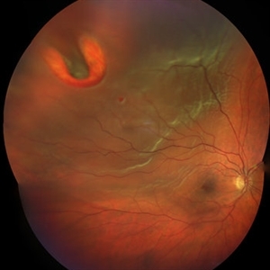
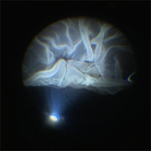
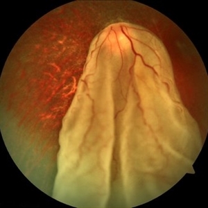 When the Curtain Falls
Jun 12 2021 by Shyamal K Dwivedi, MD
52-year-old male presented with sudden painless vision drop. Giant retinal tear discovered which was about 270 degrees anchored at the disc. Title courtesy: Dr.Aditya Sudhalkar
Photographer: Dr.Aditya Sudhalkar
Imaging device: Zeiss
Condition/keywords: giant retinal tear
-
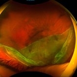
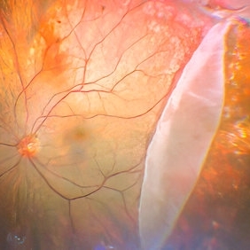
 Giant Retinal Tear
Feb 20 2024 by Soobien Lee
Optos color fundus photograph of a 40-year-old caucasian male who is a UFC fighter with a total retinal detachment in his right eye secondary to a giant retinal tear from 10 o'clock to 2 o'clock.
Photographer: Trinity Wolf, Elman Retina Group
Imaging device: Optos Ultra-Widefield Imaging
Condition/keywords: giant retinal tear, optos, Retinal Detachment, Retinal tear with detachment, trauma
-
 Giant retinal Tear
Apr 26 2022 by Jeffrey Barker
Giant retinal Tear
Photographer: Jeffrey P. Barker B.S.
Condition/keywords: retinal tear
-
 Giant Retinal Tear
May 15 2014 by Manish Nagpal, MD, FRCS (UK), FASRS
Patient presenting with a acute loss of vision with a giant retinal tear.
Photographer: pooja barot, Optometrist, Retina Foundation, Ahmedabad
Condition/keywords: giant retinal tear
-
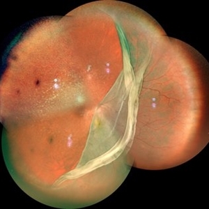
 Giant Retinal Tear
May 27 2020 by Jamin S. Brown, MD
Fundus photo montage of 55-year-old male with retinal detachment and giant retinal tear.
Photographer: Stefanie Palmer CRA, Retina-Vitreous Surgeons of CNY
Condition/keywords: giant retinal tear
-
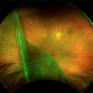
 Giant Retinal Tear
Aug 12 2021 by Stefanie Palmer
Giant Retinal Tear of the Right eye.
Photographer: Stefanie Palmer, CRA
Condition/keywords: giant retinal tear
-
 Giant Retinal Tear with Choroidal Detachment
Jun 12 2024 by Anand Temkar
Intra operative still of a 34 year old male showing Giant Retinal Tear with Choroidal Detachment.
Photographer: Dr.Anand Temkar- Retina Foundation, Ahmedabad
Condition/keywords: choroidal detachment, giant retinal tear
-
 PFO Bubbles
Feb 25 2025 by Parnian Arjmand, MD, MSc, FRCSC, DABO
Post operative day 7 after repair of an RD secondary to a giant retinal tear with temporary PFO tamponade.
Condition/keywords: GRT, PFO
-
 Retinal Detachment with Giant Retinal Tear
Mar 9 2013 by Young-Gyun Kim, MD
Fundus photograph of a 45-year-old man with retinal detachment and giant retinal tear.
Photographer: Shin Ji-Young, Eulji university, Seoul
Imaging device: Topcon TRC 50 EX
Condition/keywords: retinal tear
-
 Rhegmatogenous Retinal Detachment
Mar 3 2021 by Patrik Rajs
A 51-year-old female patient presented with inferior nasal scotoma and 5/10 vision in the right eye due to a retinal detachment with a giant retinal horseshoe tear.
Photographer: Patrik Rajs, EYE CLINIC of Jan Evangelista Purkyne University and Masaryk Hospital, Czech Republic, Ústí nad Labem
Imaging device: Clarus 700
Condition/keywords: giant retinal tear
-
Flattening of Giant retinal tear
Oct 24 2022 by Manish Nagpal, MD, FRCS (UK), FASRS
This video highlights the step of eversion of the giant retinal tear flap and flattening of retina using heavy PFCL liquid
Photographer: Manish Nagpal
Condition/keywords: flap, giant retinal tear, GRT, PFCL, video, vitrectomy
-
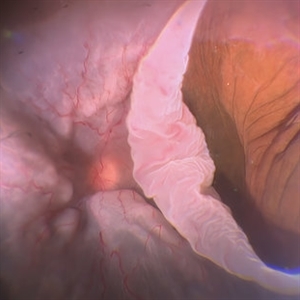
 Giant Retinal Tear
Jan 11 2022 by Manish Nagpal, MD, FRCS (UK), FASRS
Intraoperative photo a temporal giant retinal tear with everted flap and some laser marks noted on the bare choroid from previous barrage attempt elsewhere.
Photographer: Manish Nagpal, Retina Foundation, Ahmedabad, India
Imaging device: Sony PMW -10 MD surgical camera
Condition/keywords: giant retinal tear
-
 Giant Retinal Tear
Apr 1 2016 by Nichole Lewis
Giant retinal tear montaged on Anterior Segment due to the Detachment being very bullous.
Photographer: Nichole Lewis - Pennsylvania Retina Specialists, Camp Hill, PA
Condition/keywords: giant retinal tear, retinal tear
-
 Giant Retinal Tear
Jan 8 2017 by Manish Nagpal, MD, FRCS (UK), FASRS
Fundus photo of a patient complaining of floaters and a curtain like field defect.
Photographer: pravin jain
Condition/keywords: giant retinal tear
-
 Giant Retinal Tear
Apr 21 2022 by Vaidehi Sathaye
Fundus photograph of a 16 year old male with a Rhegmatogenous Retinal Detachment secondary to a Giant Retinal Tear in the right eye.
Photographer: Dr. Vaidehi Sathaye, Retina Foundation
Condition/keywords: giant retinal tear
-
 Giant Retinal Tear
Apr 19 2022 by Thais Bastos
A 44-year-old female patient with sudden loss of visual acuity in her left eye. Note retinal detachment with giant retinal tear with the retina folded over itself.
Photographer: Thaís Azeredo Bastos - HCRP-USP, Brazil
Imaging device: Optos California
Condition/keywords: giant retinal tear
-
 Giant Retinal Tear Slide 1
Oct 22 2012 by Ronald C. Gentile, MD
Acute loss of vision in a myopic man with flashes and floaters in the right eye. The giant retinal tear is flapped over with the macula detached. The undersurface of the retina can be seen temporally.
Photographer: The New York Eye & Ear Infirmary Department of Medical Imaging
Condition/keywords: retinal tear, vitrectomy
-
 Giant Tear and Vitreous Abnormalities in Stickler Syndrome
Feb 12 2021 by Anfisa Ayalon, MD
Fundus photograph of a 16-year-old male with Stickler Syndrome and giant tear rhegmatogenous retinal detachment. Note multiple vitreous veils and bands.
Photographer: Anfisa Ayalon, MD., Meir Medical Center, Kfar Saba, Israel.
Imaging device: California, Optos
Condition/keywords: empty vitreous, giant retinal tear, Stickler Syndrome, vitreous veils
-
 Intraoperative View of a Giant Retinal Tear
Dec 13 2024 by Thirumalesh Mochi Basavaraj, MD
Intraoperative view of 12 year old child with Giant retinal tear with Retinal detachment.
Photographer: Thirumalesh Mochi Basavaraj
Imaging device: Lumera Proveo 8
Condition/keywords: GIANT RETINAL TEAR, PVR, Retinal Detachment
-
 Macular Pucker in Silicon Filled Eye
May 3 2017 by Manish Nagpal, MD, FRCS (UK), FASRS
Patient operated for retinal detachment with giant retinal tear with silicon oil a month back had come for a follow up.. his retina was well attached but he had developed a macular pucker under silicon oil.
Photographer: pooja barot
Condition/keywords: macular pucker, proliferative vitreoretinopathy (PVR), silicone oil
-
 Macular Pucker Status Post Vitrectomy for GRT
Jan 4 2017 by Manish Nagpal, MD, FRCS (UK), FASRS
Macular pucker forming post 2 months of vitrectomy done for giant retinal tear.
Photographer: Pooja Barot
Condition/keywords: giant retinal tear, macular pucker, vitrectomy
-
 Retinal Detachment with Giant Retinal Tear
Aug 9 2024 by Aditya S Kelkar, MS, FRCS, FASRS,FRCOphth
Fundus photograph of an 12-year-old boy with a Retinal detachment with Giant retinal tear of acute onset.
Photographer: Sakshi Naik, National Institute of Ophthalmology
Imaging device: Optos Daytona
Condition/keywords: giant retinal tear, pediatric retina, Retina detachment
-
 Retinal Fold
Sep 26 2023 by Mauricio Bayram-Suverza, MD
A 38-year-old man underwent vitrectomy in the left eye due to a giant tear in the upper retina. SF6 gas was used as endotamponade. During the post-surgical check-up, it was identified that the patient developed a full-thickness retinal fold due to retinal slippage during fluid-air exchange. As the fold was away from the macular area, it was decided to observe the patient. Three weeks after the surgery, his best-corrected visual acuity was 20/30.
Photographer: Mauricio Bayram-Suverza, Fundación Hospital Nuestra Señora de la Luz
Imaging device: TRC-50DX
Condition/keywords: giant retinal tear, retina surgery complications, Retinal slippage, vitreoretinal surgery
-
 Retinal Tack
Oct 11 2012 by Michael P. Kelly, FOPS
This is a retinal fundus photograph I took in 1987 while working with Howard Schatz, MD and H. Richard McDonald, MD, when retinal tacks were used to repair giant retinal tears. I purposely underexposed the retina because the retinal tack is so highly relective.
Photographer: Michael P. Kelly, FOPS Director, Duke Eye Center Labs, Duke University Hospital
Condition/keywords: retinal tacks, retinal tear
-
 Silicone Oil
Jan 3 2013 by Wilfredo C. Lara, MD
Status post giant retinal tear repair with use of silicone oil.
Condition/keywords: silicone oil
 Loading…