Search results (59 results)
-
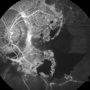
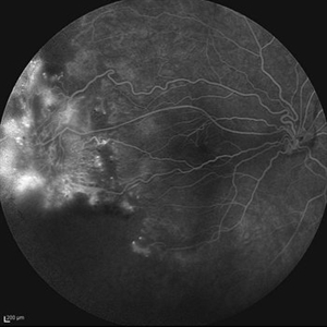
 Advanced PDR RE FFA
Aug 31 2014 by Neha Goel, MS DNB FRCS (Glasg)
Fluorescein angiogram of the right eye.
Photographer: Neha Goel
Imaging device: Zeiss Visucam
Condition/keywords: fibrovascular proliferation, ischaemic diabetic maculopathy, proliferative diabetic retinopathy (PDR)
-
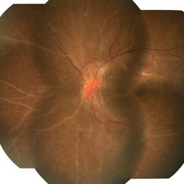
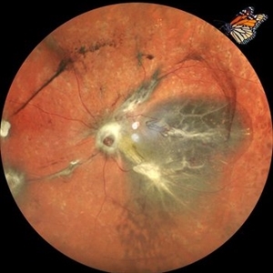
 Advanced PDR-RE
Aug 31 2014 by Neha Goel, MS DNB FRCS (Glasg)
Fundus photograph of the right eye of a 50-year-old diabetic male.
Photographer: Neha Goel
Imaging device: Zeiss Visucam
Condition/keywords: fibrovascular proliferation, ischaemic diabetic maculopathy, proliferative diabetic retinopathy (PDR)
-
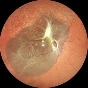
 Advanced Proliferative Diabetic Retinopathy With Fibrovascular Proliferation
Jan 4 2019 by Isha Agarwalla
A 29-year-old female with a long-standing history of diabetes mellitus presented with a fibrovascular membrane (FVM) at the viteroretinal interface due to underlying inflammation and angiogenesis induced by ischemia. FVM involved the disc and extended towards the superior and inferior arcades along with extensive capillary drop out areas due to micro aneurysms.
Condition/keywords: fibrovascular proliferation, proliferative diabetic retinopathy (PDR)
-
 The Bullet Ridden Retina
Feb 17 2024 by SHISHIR VERGHESE, MS, FVRS, FAICO (Retina)
Fundus image obtained of a case of lasered branch retinal vein occlusion (BRVO) with fibrovascular proliferation (FVP) where the laser marks have given way to multiple small retinal holes due to traction from the same.
Photographer: DIVYA SHAJI
Imaging device: NIDEK MIRANTE
Condition/keywords: BRVO, chronic retinal detachment
-
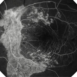
 Advanced PDR Left Eye FFA
Aug 31 2014 by Neha Goel, MS DNB FRCS (Glasg)
Fluorescein angiogram of the left eye.
Photographer: Neha Goel
Imaging device: Zeiss Visucam
Condition/keywords: fibrovascular proliferation, ischaemic diabetic maculopathy, proliferative diabetic retinopathy (PDR)
-
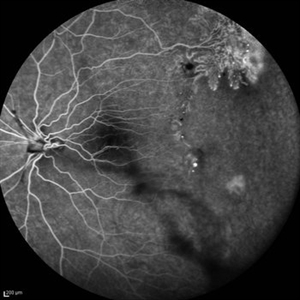 Peripheral Nonperfusion and Optic Disc Hypoplasia
Oct 13 2012 by Hamid Ahmadieh, MD
FA image of the left eye of a 24-year-old woman with peripheral retinal nonperfusion and fibrovascular proliferation associated with optic disc hypoplasia; visual acuity of 20/50. Her brother and sister were also involved with the same ocular disorder.
Photographer: Hamid Ahmadieh, MD, Ophthalmic Research Center, Labbafinejad Medical Center, Shahid Beheshti University of Medical Sciences
Imaging device: Heidelberg Spectralis
Condition/keywords: fibrovascular proliferation, optic disc hypoplasia, peripheral retinal nonperfusion
-
 Peripheral Nonperfusion and Optic Disc Hypoplasia
Oct 13 2012 by Hamid Ahmadieh, MD
Late FA image of the right eye of a 24-year-old woman with peripheral retinal nonperfusion and fibrovascular proliferation associated with optic disc hypoplasia; visual acuity of 20/50. Her brother and sister were also involved with the same ocular disorder.
Photographer: Hamid Ahmadieh, MD, Ophthalmic Research Center, Labbafinejad Medical Center, Shahid Beheshti University of Medical Sciences
Imaging device: Heidelberg Spectralis
Condition/keywords: fibrovascular proliferation, optic disc hypoplasia, peripheral retinal nonperfusion
-
 Fibrovascular Proliferation
Jul 28 2018 by Juan Romo-Aguas
Fundus photograph of a patient right eye with a fibrovascular proliferation secondary to proliferative diabetic retinopathy.
Photographer: Juan Romo-Aguas, Asociacio´n para Evitar la Ceguera en Me´xico, Hospital ‘‘Dr. Luis Sa´nchez Bulnes’’ I.A.P., Mexico
Imaging device: Optos Daytona Ultra-widefield Retinal Imaging
Condition/keywords: proliferative diabetic retinopathy (PDR)
-
 Proliferative Diabetic Retinopathy with Tractional Retinal Detachment
Mar 15 2018 by awaneesh m upadhyay, MBBS, DNB
Right eye fundus photo of a 57-year-old male with proliferative diabetic retinopathy and tractional retinal detachment.
Photographer: Awaneesh Upadhyay
Condition/keywords: fibrovascular proliferation, proliferative diabetic retinopathy (PDR), tractional retinal detachment
-
 Advanced PDR Left Eye
Aug 31 2014 by Neha Goel, MS DNB FRCS (Glasg)
Fundus photograph of the left eye.
Photographer: Neha Goel
Imaging device: Zeiss Visucam
Condition/keywords: fibrovascular proliferation, ischaemic diabetic maculopathy, proliferative diabetic retinopathy (PDR)
-
 Advanced Proliferative Diabetic Retinopathy With Fibrovascular Proliferation
Jan 4 2019 by Isha Agarwalla
A 29-year-old female with a long-standing history of diabetes mellitus presented with a fibrovascular membrane(FVM) at the viteroretinal interface due to underlying inflammation and angiogenesis induced by ischemia. FVM involved the disc and extended towards the superior and inferior arcades along with extensive capillary drop out areas due to micro aneurysms.
Condition/keywords: fibrovascular proliferation, fluorescein angiogram (FA), proliferative diabetic retinopathy (PDR)
-
---thumb.jpg/image-square;max$300,300.ImageHandler) Anterior Hyaloid Fibrovascular Proliferation
Anterior Hyaloid Fibrovascular Proliferation
Feb 13 2013 by From the Collections of Thomas M. Aaberg, MD and Thomas M. Aaberg Jr., MD
Histopathology neovascularization.
Condition/keywords: fibrovascular proliferation, histopathology, hyaloid, neovascularization (NV)
-
 Before and After Vitrectomy
Nov 17 2023 by Bradley T. Smith, MD, FASRS
Middle age male diabetic retinopathy and resolving exudate following repair of tractional detachment with membrane peeling.
Condition/keywords: coats-like response, Diabetes, fibrotic neovascularization, fibrovascular proliferation, pars plana vitrectomy (PPV), proliferative diabetic retinopathy (PDR), tractional retinal detachment
-
 Bilateral Severe PDR
May 3 2015 by Mallika Goyal, MD
Right fundus of a young lady with PDR shows vitreous haemorrhage, fibrovascular proliferation from disc with inferior TRD. Fellow eye has PDR with NVD.
Photographer: Mallika Goyal, MD, Apollo Health City, Jubilee Hills, Hyderabad
Condition/keywords: proliferative diabetic retinopathy (PDR)
-
 Combined Retinal Detachment With a Butterfly Shaped Configuration
Mar 13 2025 by S. Natarajan, MD, FASRS, FRCS (GLASGOW) , FICO, D.Sc, FELA
A 46 year old female presented to us with diminished vision in both the eyes. Her blood glucose levels were deranged. She had bilateral proliferative diabetic retinopathy and pan retinal photocoagulation was done elsewhere. Left eye showed a combined retinal detachment with fibrovascular proliferation on the disc and along inferior arcade with a convex configuration of retinal detachment. Patient was planned for surgical intervention. The image shows a butterfly like configuration of combined retinal detachment with the subretinal fluid pocket appearing like the wings of the butterfly.
Photographer: ASHWINI SUTAR ADITYA JYOT EYE HOSPITAL
Imaging device: Mirante ( PLEASE SELECT COVER PAGE )
Condition/keywords: retinal detachment with a butterfly shaped
-
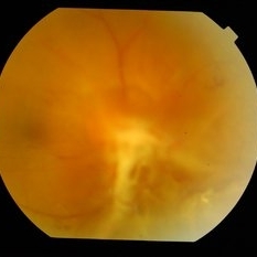
 Diabetic Proliferative Retinopathy
Dec 1 2019 by Lucas Zago Ribeiro, MD
Fundus photograph of 75-year-old man with diabetic proliferative retinopathy with fibrovascular proliferation over the optic disc.
Photographer: Lucas Zago Ribeiro, Federal University of São Paulo
Imaging device: Zeiss Visucam 524
Condition/keywords: diabetic retinopathy, fibrovascular proliferation, neovascularization (NV)
-
 Diabetic Tractional Retinal Detachment
Jan 6 2025 by Kavitha Duraipandi, MD DNB FICO FRCS
55 year old patient , with poor metabolic control , came with right eye gradual loss of vision. Patient had partial inadequate retinal laser in the past. Funds examination showed fibrovascular proliferation over the arcades with tractional retinal detachment. Patient under went right eye Pars plans vitrectomy with endo laser with silicone oil injection. patient was given pre op anti VEGF injection.
Condition/keywords: TRD
-
 Fibrotic Tractional Membrane in ROP Stage 5
Nov 7 2013 by Maria Ana Martinez-Castellanos, MD
Stage 5 retinopathy of prematurity in a 6 month old baby.
Photographer: Maria A. Martinez-Castellanos. Asociacion para Evitar la Ceguera en Mexico
Imaging device: RetCam II
Condition/keywords: fibrous proliferation, fibrovascular proliferation, retinopathy of prematurity (ROP)
-
---thumb.jpg/image-square;max$300,300.ImageHandler) Fibrovascular Proliferation
Fibrovascular Proliferation
Feb 13 2013 by From the Collections of Thomas M. Aaberg, MD and Thomas M. Aaberg Jr., MD
Disc involvement, neovascularization, tractional detachment.
Condition/keywords: disc, fibrovascular proliferation, neovascularization (NV), tractional retinal detachment
-
---thumb.jpg/image-square;max$300,300.ImageHandler) Fibrovascular Proliferation
Fibrovascular Proliferation
Feb 13 2013 by From the Collections of Thomas M. Aaberg, MD and Thomas M. Aaberg Jr., MD
Disc involvement, neovascularization, tractional detachment.
Condition/keywords: disc, fibrovascular proliferation, neovascularization (NV)
-
---thumb.jpg/image-square;max$300,300.ImageHandler) Fibrovascular Proliferation
Fibrovascular Proliferation
Feb 13 2013 by From the Collections of Thomas M. Aaberg, MD and Thomas M. Aaberg Jr., MD
Neovascularization, fibrous proliferation, intraretinal hemorrhage.
Condition/keywords: fibrous proliferation, intraretinal hemorrhage, neovascularization (NV)
-
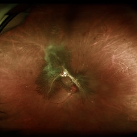
 FLORID Type PDR OS
Mar 17 2020 by Deepak Bhojwani, MS
Left eye montage fundus image of a diabetic patient with FLORID fibrovascular proliferation around optic disc .
Photographer: DEEPAK BHOJWANI
Condition/keywords: fibrovascular proliferation, florid type PDR, proliferative diabetic retinopathy (PDR)
-
 Lower Venous Branch Occlusion With Fibrovascular Proliferation
Oct 25 2016 by Daniel Rojas Abatte
Fundus photograph of an 58-year-old female patient with fibrovascular proliferation product of an occlusion of ancient venous branch.
Photographer: Daniel Rojas A
Imaging device: TRC-50DX Topcon
Condition/keywords: fibrovascular proliferation, fundus photograph
 Loading…