Search results (103 results)
-
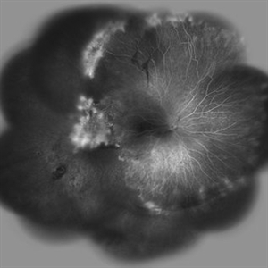
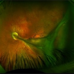
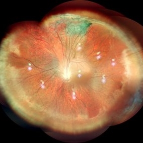
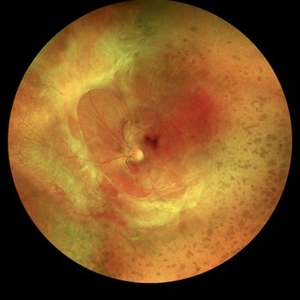 Familial Exudative Vitreoretinopathy
Jan 21 2019 by Netan Choudhry, MD, FRCS(C) FASRS
Widefield montage pseudocolor image of a 35-year-old woman with prior history of FEVR.
Photographer: Carmelina Timboli, Vitreous Retina Macula Specialists of Toronto
Imaging device: Optos California (Optos PLC, Edinburgh, UK)
Condition/keywords: familial exudative vitreoretinopathy (FEVR)
-
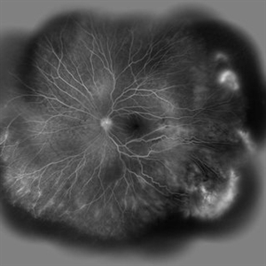
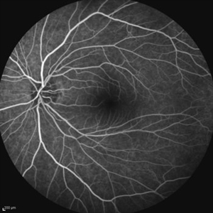
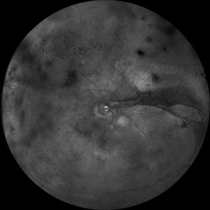
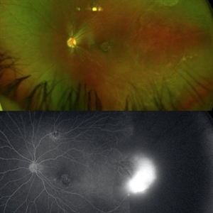 Familial Exudative Vitreoretinopathy
Jan 21 2021 by Gabriel Costa Andrade, PhD
Fundus photograph and angiography of an 4-year-old boy with Stage 2 FEVR.
Photographer: Gabriel Andrade
Condition/keywords: familial exudative vitreoretinopathy (FEVR)
-
 Familial Exudative Vitreoretinopathy
Feb 4 2022 by Naresh Babu Kannan, MS, FNB(V R),MBA (H R),FASRS,.
Wide field fundus photograph of a 20-year-old woman with familial exudative vitreoretinopathy showing temporal avascular retinal periphery. BCVA OD 20/40.
Photographer: Mrs. Bharathi, Aravind Eye Hospital, Madurai
Imaging device: Zeiss Clarus
Condition/keywords: familial exudative vitreoretinopathy (FEVR), pediatric retinal vascular diseases, temporal avascular retina
-
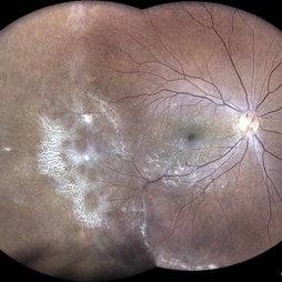
 Familial Exudative Vitreoretinopathy
Nov 25 2022 by Aditya S Kelkar, MS, FRCS, FASRS,FRCOphth
Colour fundus photograph of the right eye of a 56-year-old lady showing lasered FEVR with epiretinal membrane and vitreous band.
Photographer: Dr. Pranali Surawase. National Institute of Ophthalmology, Pune, Maharashtra, India
Imaging device: Zeiss Clarus 500
Condition/keywords: ERM, familial exudative vitreoretinopathy (FEVR), laser photocoagulation
-
 Familial Exudative Vitreoretinopathy
Jan 28 2023 by Krushna Gopal Panda
Fundus photograph of a six month-old baby with Familial Exudative Vitreoretinopathy
Photographer: Krushna Gopal Panda
Imaging device: Optos california
Condition/keywords: familial exudative vitreoretinopathy (FEVR)
-
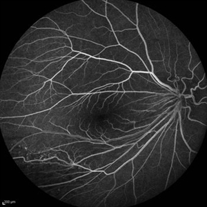 Familial Exudative Vitreoretinopathy (FEVR)
Oct 13 2012 by Hamid Ahmadieh, MD
Early FA image of the right eye of a 24-year-old woman with FEVR and visual acuity of 20/25.
Photographer: Hamid Ahmadieh, MD, Ophthalmic Research Center, Labbafinejad Medical Center, Shahid Beheshti University of Medical Sciences
Imaging device: Heidelberg Spectralis
Condition/keywords: familial exudative vitreoretinopathy (FEVR)
-
 FEVR+ Optic Disc Hypoplasia
Oct 13 2012 by Hamid Ahmadieh, MD
FA image of the right eye of a 24-year-old woman with FEVR and optic disc hypoplasia; visual acuity of 20/50. Her brother and sister were also involved with the same ocular disorder.
Photographer: Hamid Ahmadieh, MD, Ophthalmic Research Center, Labbafinejad Medical Center, Shahid Beheshti University of Medical Sciences
Imaging device: Heidelberg Spectralis
Condition/keywords: familial exudative vitreoretinopathy (FEVR), optic disc hypoplasia
-
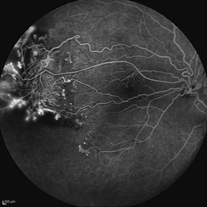
 FEVR+ Optic Disc Hypoplasia
Oct 13 2012 by Hamid Ahmadieh, MD
Wide field FA image of the right eye of a 24-year-old woman with FEVR and optic disc hypoplasia; visual acuity of 20/50. Her brother and sister were also involved with the same ocular disorder.
Photographer: Hamid Ahmadieh, MD, Ophthalmic Research Center, Labbafinejad Medical Center, Shahid Beheshti University of Medical Sciences
Imaging device: Heidelberg Spectralis
Condition/keywords: familial exudative vitreoretinopathy (FEVR), optic disc hypoplasia
-
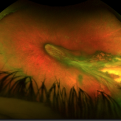
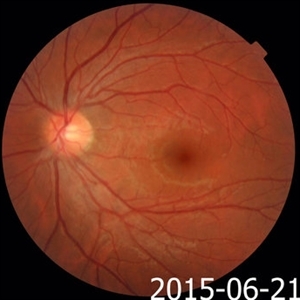
 Familial Exudative Vitreoretinopathy
Mar 13 2013 by Everton L. Arrindell, MD
Familial Exudative Vitreoretinopathy (FEVR).
Photographer: Alecia Camp, CRA - Tennessee Retina - Nashville, TN
Condition/keywords: familial exudative vitreoretinopathy (FEVR)
-
 FEVR
FEVR
May 8 2014 by S. Natarajan, MD, FASRS, FRCS (GLASGOW) , FICO, D.Sc, FELA
Fundus photograph of a 8-year-old female with C/O DM/VN OS showing vitreous traction band dragging of macular and disc OD showed retinal detachment
Photographer: ADITYA JYOT EYE HOSPITAL,MUMBAI INDIA
Condition/keywords: familial exudative vitreoretinopathy (FEVR)
-
 Familial Exudative Vitreoretinopathy
Aug 18 2021 by Samuel Dada
Ultra-widefield optos image of a 40-year with Familial Exudative Vitreoretinopathy, affecting his left eye. Patient born at 38 weeks. No NICU time. Has had genetic testing to determine cause of blindness. Physician suspects FEVR and will carry out further testing. Patient uses a 200x or 600x magnifying lens to view and focus on objects at a distance. Patient's vision on initial visit was 20/70.
Photographer: Samuel Dada
Imaging device: Optos California
Condition/keywords: dysplastic excavation, familial exudative vitreoretinopathy (FEVR), fundus photograph, left eye, Optos, pseudocolor, ultra-wide field imaging
-
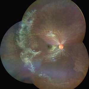
 Familial Exudative Vitreoretinopathy (FEVR)
Apr 24 2021 by Alexandre Grandinetti, MD, PhD
6-year-old girl with retinal folds on both eyes secondary to FEVR.
Photographer: Corina Szrek
Imaging device: Optos California
Condition/keywords: familial exudative vitreoretinopathy (FEVR)
-
 Familial Exudative Vitreoretinopathy (FEVR)
Oct 13 2012 by Hamid Ahmadieh, MD
Wide field FA image of the Left eye of a 24-year-old woman with FEVR and visual acuity of 20/30.
Photographer: Hamid Ahmadieh, MD, Ophthalmic Research Center, Labbafinejad Medical Center, Shahid Beheshti University of Medical Sciences
Imaging device: Heidelberg Spectralis
Condition/keywords: familial exudative vitreoretinopathy (FEVR)
-
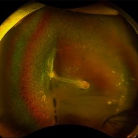
 FEVR with Knife-Like Fold
Jan 11 2014 by Caesar K. Luo, MD, FASRS
Retcam photograph of child with FEVR.
Photographer: Caesar Luo, Progressive Vision Institute, PA
Imaging device: RetCam
Condition/keywords: familial exudative vitreoretinopathy (FEVR)
-
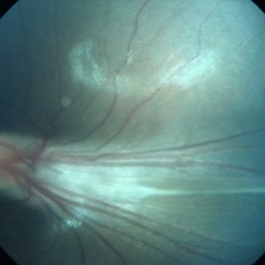
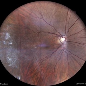
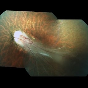 Familial Exudative Vitreoretinopathy
Jan 11 2018 by S. Natarajan, MD, FASRS, FRCS (GLASGOW) , FICO, D.Sc, FELA
Fundus Photograph of 25-year-old lady with Familial Exudative Vitreoretinopathy : Left eye showing pale optic disc with disc drag and sheathing of vessels.
Photographer: Miss Ashwini Borde
Imaging device: Carl Zeiss 450 plus IR
Condition/keywords: familial exudative vitreoretinopathy (FEVR)
-
 A Classic Case of Retinal Ora Serrata Imaging
Jan 16 2025 by yuan duo
A 5-year-old girl, born full-term with no history of systemic disease, presented with poor vision since early childhood and underwent fundus examination. Anterior segments of both eyes showed no significant abnormalities. Fundus examination revealed retinal folds extending from the optic disc to the temporal peripheral retina, with blood vessels coursing through the folds (A, B). Avascular zones were observed in the peripheral retina, and the ora serrata’s boundaries were clearly visible, displaying dentate processes and bays (C, D). Retinal pigmentation was evident. Genetic testing confirmed the final diagnosis of bilateral Familial Exudative Vitreoretinopathy (FEVR).
Condition/keywords: Retinal Ora Serrata
-
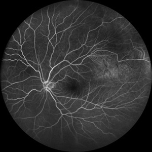
 Asymptomatic Eye in FEVR
Jul 7 2015 by Hamid Ahmadieh, MD
FA image of the asymptomatic left eye of a 28-year-old man with total RD secondary to advanced FEVR in his right eye. Notice straightening of the retinal vessels.
Photographer: Soulmaz Shahmohammad, Negah Eye Center, Tehran, Iran
Imaging device: Specteralis
Condition/keywords: asymptomatic, familial exudative vitreoretinopathy (FEVR)
-
 Asymptomatic Eye in FEVR
Jul 7 2015 by Hamid Ahmadieh, MD
Color fundus photograph of the asymptomatic eye of a patient with FEVR. Notice straightening of the retinal vessels.
Photographer: Soulmaz Shahmohammad, Negah Eye Center, Tehran, Iran
Condition/keywords: color fundus photograph, familial exudative vitreoretinopathy (FEVR)
-
 FA OS FEVR
Aug 15 2021 by ASRS Staff
15 year-old male presented with squinting and diminution of vision in OD. His vision was finger count at 2 meters in OD and 6/6 in OS. On posterior segment examination, peripheral capillary agenesis and aberrant circumferential vessels were present in both eyes.
Imaging device: Nidek Mirante
Condition/keywords: familial exudative vitreoretinopathy (FEVR), fluorescein angiogram (FA)
-
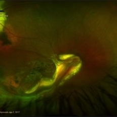 Familial Exsudative Vitreoretinopathy (FEVR)
Aug 4 2017 by Alexandre Grandinetti, MD, PhD
Fundus photograph of a 16-year-old monocular girl with FEVR.
Photographer: Corina Shrzek
Imaging device: P200DTX California - Optos
Condition/keywords: familial exudative vitreoretinopathy (FEVR)
-
 Familial Exudative Vitreo-retinopathy
Jul 6 2021 by Akansha Sharma
Color photo montage of 21-year-old male with familial exudative vitreoretinopathy in an amblyopic eye.
Photographer: Dr. Akansha Sharma-Retina Foundation, Ahmedabad
Condition/keywords: familial exudative vitreoretinopathy (FEVR), retinal ischemia
-
 Familial Exudative Vitreo-retinopathy
Jul 6 2021 by Akansha Sharma
Infra-red capture of 5-year-old male with familial exudative vitro-retinopathy with disc pallor.
Photographer: Dr. Akansha Sharma-Retina Foundation, Ahmedabad
Condition/keywords: familial exudative vitreoretinopathy (FEVR), retinal ischemia
-
 Familial Exudative Vitreo-Retinopathy
May 8 2023 by Akansha Sharma
Colour fundus photograph of a 28 year old female with familial exudative vitreoretinopathy
Photographer: Dr. Urmil Shah, Dr. Denish Patel, Dr. Akansha Sharma, Bharati Eye Clinic, Ahmedabad, Gujarat
Condition/keywords: familial exudative vitreoretinopathy (FEVR)
-
 Familial Exudative Vitreo-Retinopathy
Jan 30 2024 by Akansha Sharma
Colour fundus photograph of a 19 year old male with both eyes familial exudative vitreo-retinopathy. Right eye shows peripheral avasular areas.
Photographer: Dr. Akansha Sharma, Bharati Eye Hospital
Condition/keywords: familial exudative vitreoretinopathy (FEVR), REGRESSED ROP
-
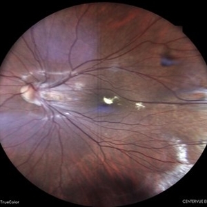 Familial Exudative Vitreo-Retinopathy
Jan 30 2024 by Akansha Sharma
Colour fundus photograph of a 19 year old male with both eyes familial exudative vitreo-retinopathy. Left eye shows aggregation of hard exudates over the fovea.
Photographer: Dr. Akansha Sharma, Bharati Eye Hospital
Condition/keywords: familial exudative vitreoretinopathy (FEVR), REGRESSED ROP
 Loading…