Search results (11 results)
-
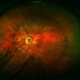
 Degenerative Myopia
Aug 23 2012 by Gabriela Lopezcarasa Hernandez, MD
Photographer: Gabriela Lopezcarasa Hernandez, Macula Retina Consultores
Imaging device: Heidelberg Spectralis
Condition/keywords: degenerative myopia
-
 Color Fundus Photograph of Myope With PVD and Staphyloma
Jun 11 2016 by Philip J. Polkinghorne, MD
Color photograph of patient with PVD and staphyloma.
Imaging device: Optos
Condition/keywords: degenerative myopia, peripheral vascular disease (PVD), staphyloma
-
 Degenerative Myopia
Apr 12 2023 by Ahmed Abbas Hashmi, OD
Right eye Fundus photograph of a 61-year-old female with pathological myopia.
Condition/keywords: chorioretinal atrophy, high myopia, pathologic myopia
-
 Degenerative Myopia
Apr 21 2024 by César Adrián Gómez Valdivia, MD
Degenerative Myopia
Photographer: Erika Paulina Ornelas Cazares
Imaging device: Topcon TRC-50 DX
Condition/keywords: degenerative myopia, myopia
-
 Macular Dystrophy vs Myopic Degeneration
Dec 22 2023 by Virginia Gebhart
35 year old female with myopic degeneration (-18.00 OU). BCVA 20/100 OU. RPE atrophy present in both eyes, but no significant chorioretinal atrophy. OCT not consistent with degenerative myopia due to dome shape appearance rather than posterior bowing. Possible macular dystrophy over degeneration. Will observe
Photographer: Virginia Gebhart
Imaging device: Topcon
Condition/keywords: Macular Dystrophy, myopic degeneration
-
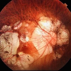
 Macular Hole Retinal Detachment Over a Posterior Staphyloma
Dec 31 2016 by Linda A Cernichiaro- Espinosa, MD
Macular hole retinal detachment over a posterior staphyloma of pathologic myopia.
Photographer: Linda A Cernichiaro
Imaging device: Optos
Condition/keywords: degenerative myopia, high myopia, macular hole, myopic eye, posterior staphyloma, vitreoretinal degeneration
-
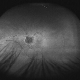 Myope With Staphyloma and Vitreous Detachment
Jun 11 2016 by Philip J. Polkinghorne, MD
Fundus autofluorescence of a myope with PVD and staphyloma.
Imaging device: Optos FAF
Condition/keywords: degenerative myopia, myopia, staphyloma
-
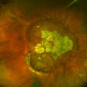 Posterior staphyloma
Dec 20 2023 by Roger A. Goldberg, MD, MBA
Fundus photo of an 85-year-old woman with degenerative myopia and a large posterior staphyloma
Photographer: Mohan Zhou, Bay Area Retina Associates, Walnut Creek, CA
Imaging device: Optos
Condition/keywords: degenerative myopia, high myopia, posterior staphyloma
-
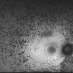 Retinitis Pigmentosa - Autofluorescence OD
Jun 18 2018 by Hosam Attia, MD
Ultra-wide fundus auto-fluorescence photograph of a 38-year-old African, American female with degenerative myopia, unilateral RP variant, depicting extensive mid-peripheral bone spicules hypo-autofluorescence, extending further into the periphery w/ relative sparing of the macula OD VF 30-V showed severe peripheral constriction OD, enlarged BS OS and OCT showed severe ellipsoid zone degeneration with saucerization and cystoid macular degeneration with no obvious late macular leakage on FA (Both, not shown)
Imaging device: Optos California
Condition/keywords: bone spicule, peripheral bone spicules, retinitis pigmentosa
-
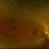 Retinitis Pigmentosa - Color OD
Jun 18 2018 by Hosam Attia, MD
Pseudo-color ultra-wide fundus photograph of a 38-year-old African American female with degenerative myopia and Unilateral RP variant, depicting extensive mid-peripheral bone spicules, extending further into the periphery, with relative sparing of the macula OD. VF 30-V showed severe peripheral constriction OD, enlarged BS OS and OCT showed severe ellipsoid zone degeneration with saucerization and cystoid macular degeneration with no obvious late macular leakage on FA (Both, not shown)
Imaging device: Optos California
Condition/keywords: bone spicule, peripheral bone spicules, retinitis pigmentosa
-
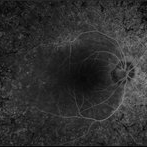 Retinitis Pigmentosa - Fluorescein Angiogram OD
Jun 18 2018 by Hosam Attia, MD
Ultra-wide fluorescein angiogram of a 38-year-old African, American female with degenerative myopia, Unilateral RP variant, depicting abnormal fluorescence pattern with extensive mid-peripheral bone spicules hypofluorescence, extending further into the periphery w/ relative sparing of the macula OD. VF 30-V showed severe peripheral constriction OD, enlarged BS OS & OCT showed severe ellipsoid zone degeneration with saucerization and cystoid macular degeneration w/ No obvious late macular leakage on FA (Both, not shown)
Imaging device: Optos California
Condition/keywords: bone spicule, peripheral bone spicules, retinitis pigmentosa
 Loading…