 Initializing download.
-
 By Hosam Attia, MD
- Uploaded on Jun 18, 2018.
- Last modified by Caroline Bozell on Jun 19, 2018.
- Rating
- Appears in
- Retinitis Pigmentosa
- Condition/keywords
- bone spicule, peripheral bone spicules, retinitis pigmentosa
- Imaging device
-
Scanning laser ophthalmoscope
Optos California - Description
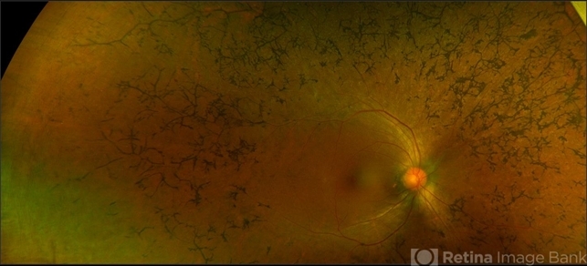
- Pseudo-color ultra-wide fundus photograph of a 38-year-old African American female with degenerative myopia and Unilateral RP variant, depicting extensive mid-peripheral bone spicules, extending further into the periphery, with relative sparing of the macula OD. VF 30-V showed severe peripheral constriction OD, enlarged BS OS and OCT showed severe ellipsoid zone degeneration with saucerization and cystoid macular degeneration with no obvious late macular leakage on FA (Both, not shown)


---thumb.jpg/image-square;max$79,0.ImageHandler "Retinitis Pigmentosa")


