Search results (23 results)
-
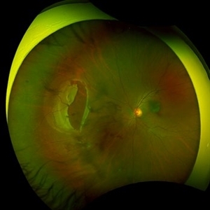
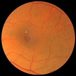
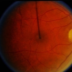
 Hemangioma of Retina
Mar 5 2025 by Virginia Gebhart
64 year old male with choroidal hemangioma in the macula and STA. Persistent IRF and new cuff of SRF compared to previous photos. BCVA CF@face. Pt has had PDT in the past with no significant improvement. Will observe closely
Photographer: Virginia Gebhart, Retina Consultants of Carolina
Imaging device: Optos California
Condition/keywords: hemangioma, inferior subretinal fluid
-
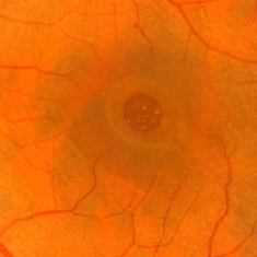
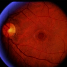
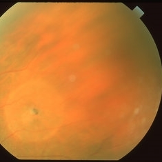
 Hemangioma of Retina (FAF)
Mar 5 2025 by Virginia Gebhart
Fundus autofluorescence of 64 year old male with choroidal hemangioma in the macula and STA. Persistent IRF and new cuff of SRF compared to previous photos. BCVA CF@face. Pt has had PDT in the past with no significant improvement. Will observe closely
Photographer: Virginia Gebhart, Retina Consultants of Carolina
Imaging device: Optos California
Condition/keywords: autofluorescence imaging, hemangioma, inferior subretinal fluid
-
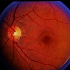
 Large Retinal Tear from a Shuttlecock Injury
Oct 11 2024 by Ahmad B. Tarabishy, MD
27 year old woman presenting with floaters and a shadow in her temporal visual field OS. Approximately one week earlier, she was injured in her left eye by a shuttlecock while playing badminton. Fundus exam reveals mild vitreous hemorrhage and a large retinal tear with a small cuff of surrounding SRF.
Photographer: Angela Rico, M.D.
Imaging device: Optos
Condition/keywords: blunt trauma, ocular trauma, retinal tear
-
 Macular Hole
Mar 29 2013 by Henry J. Kaplan, MD
Chronic macular hole with drusen like deposits and surrounding cuffing of subretinal fluid.
Condition/keywords: macular hole
-
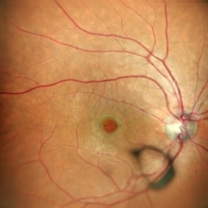
Peripheral Retinal Hole with OCT Co-localization
Sep 26 2023 by Bradley T. Smith, MD, FASRS
Peripheral asymptomatic atrophic retinal hole with OCT co localization demonstrating small cuff of sub retinal fluid. Near infrared imaging shows hyper reflectivity through hole.
Condition/keywords: atrophic hole, lattice degeneration, OCT
-
 Macular Hole
May 11 2020 by Gayathri Mohan
Color fundus photograph of a patient with macular hole along with surrounding cuff of fluid. A Weiss ring can be seen anteriorly in the vitreous.
Photographer: Gayathri Mohan, Retina Foundation
Imaging device: Mirante, Nidek
Condition/keywords: macular hole
-
 Macular Hole
Sep 27 2012 by Jeffrey G. Gross, MD, FASRS
Macular hole, chronic with fluid cuff and deposits in bed of hole.
Condition/keywords: macular hole, subretinal fluid
-
 Diffuse uveal melanoma
Dec 25 2022 by Giovanni Cuffaro, MD
Left eye of a bilateral diffuse uveal melanoma with liver metastases. Complete monosomy 3 and HLA-A*0201–positive was detected.
Photographer: Giovanni Cuffaro, Università Cattolica del Sacro Cuore, Fondazione Policlinico A. Gemelli IRCCS
Imaging device: California from Optos
Condition/keywords: Bilateral, Diffuse Choroidal Melanoma, Monosomy 3
-
 Full-thickness Macular Hole
Apr 8 2019 by Gary R. Cook, MD, FACS
Elderly white female with a Stage IV, full-thickness macular hole OS with whitish deposits visible at the base of the hole and a surrounding cuff of subretinal fluid
Imaging device: Topcon VT-50
Condition/keywords: full thickness macular hole, macular hole
-
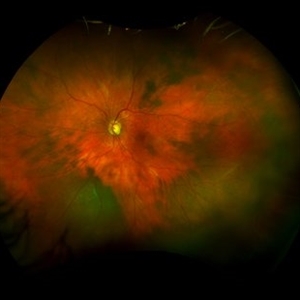
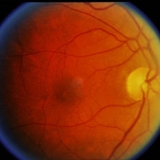
 Large Retinal Tear from a Shuttlecock Injury
Oct 11 2024 by Ahmad B. Tarabishy, MD
27 year old woman presenting with floaters and a shadow in her temporal visual field OS. Approximately one week earlier, she was injured in her left eye by a shuttlecock while playing badminton. Fundus exam reveals mild vitreous hemorrhage and a large retinal tear with a small cuff of surrounding SRF. This image was taken immediately following treatment with barrier laser retinopexy.
Photographer: Angela Rico, M.D.
Imaging device: Optos
Condition/keywords: blunt trauma, ocular trauma, retinal tear
-
 Macular Hole
Sep 27 2012 by Jeffrey G. Gross, MD, FASRS
Macular hole s/p 360 degree laser to fluid cuff.
Condition/keywords: macular hole, subretinal fluid
-
 Macular Hole
Sep 27 2012 by Jeffrey G. Gross, MD, FASRS
Macular hole magnified with cuff of SRF.
Condition/keywords: cuff, macular hole, subretinal fluid
-
 Macular Hole
Oct 8 2012 by Jeffrey G. Gross, MD, FASRS
Macular hole, s/p PPV, not closed, with residual subretinal gass bubble, in cuff.
Condition/keywords: cuff, macular hole
-
 Macular Hole - Large Hole With Cuff
Dec 19 2014 by David Callanan, MD
Macular hole - large hole with cuff .
Condition/keywords: macular hole
-
 Macular Hole - Large Hole With Cuff
Dec 19 2014 by David Callanan, MD
Macular hole - large hole with cuff .
Condition/keywords: macular hole
-
 Macular Hole - Large Hole With Cuff
Dec 19 2014 by David Callanan, MD
Macular hole - large hole with cuff .
Condition/keywords: macular hole
-
 Macular Hole - Large Hole With Cuff
Dec 19 2014 by David Callanan, MD
Macular hole - large hole with cuff .
Condition/keywords: macular hole
-
 Operculated Retinal Hole
Apr 8 2019 by Gary R. Cook, MD, FACS
White female with an operculated retinal hole with a small cuff of surrounding SRF; V.A. = 20/25
Imaging device: Topcon VT-50
Condition/keywords: operculated retinal hole
-
 Retinal Hole with Subclinical Detachment
Apr 8 2019 by Gary R. Cook, MD, FACS
64-year-old white female with an asymptomatic retinal hole with some pigment and a 1DD surrounding cuff of subretinal fluid; V.A. = 20/20-3.
Imaging device: Topcon VT-50
Condition/keywords: retinal hole, subclinical detachment
-
 Slide 1-22
Feb 19 2019 by Lancaster Course in Ophthalmology
Lymphocytes and macrophages around a vein ("cuffing") in the retina of a patient with endophthalmitis. (H&E stain)
Condition/keywords: endophthalmitis, lymphocytes, macrophages
-
---thumb.jpg/image-square;max$300,300.ImageHandler) Struck With Hairbrush
Struck With Hairbrush
Oct 15 2013 by Maurice F. Rabb
This 12 year old girl had perfectly normal vision in both eyes until she was struck in the right eye with a hairbrush during a bathroom scuffle with her sister. She is now 20/50 OD and 20/15 OS. The FFA was performed a few weeks earlier than the color photograph.
 Loading…