Search results (93 results)
-
---thumb.jpg/image-square;max$300,300.ImageHandler) Central Retinal Vein Occlusion
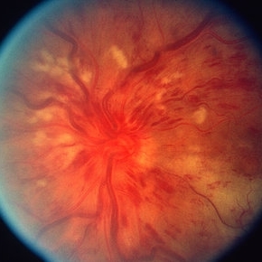
Central Retinal Vein Occlusion
Oct 30 2012 by Lihteh Wu, MD
35-year-old hypertensive man with an acute CRVO. Notice the peripapillary cotton wool spots, superficial flame shaped hemorrhages and deeper dot and blot hemorrhages in all 4 quadrants. This is the typical blood and thunder appearance of a CRVO.
Condition/keywords: central retinal vein occlusion (CRVO), cotton wool spots
-
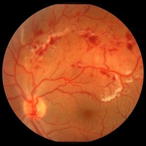
 Acute Idiopathic Occlusive Retinal Vasculitis
May 31 2014 by Hamid Ahmadieh, MD
Color fundus photograph of the right eye of a 28-year-old woman with sudden drop of vision due to acute occlusive retinal vasculitis leading to extensive nerve fiber layer infarction and retinal hemorrhages.
Photographer: Naghmeh Nozhat, Negah Eye Center, Tehran
Condition/keywords: color fundus photograph, cotton wool spots, retinal hemorrhage, retinal ischemia
-
---thumb.jpg/image-square;max$300,300.ImageHandler) Central Retinal Vein Occlusion
Central Retinal Vein Occlusion
Oct 30 2012 by Lihteh Wu, MD
35-year-old hypertensive man with an acute CRVO. Notice the peripapillary cotton wool spots, superficial flame shaped hemorrhages and deeper dot and blot hemorrhages in all 4 quadrants. This is the typical blood and thunder appearance of a CRVO.
Condition/keywords: central retinal vein occlusion (CRVO), cotton wool spots
-
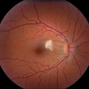
 Central Retinal Vein Occlusion associated with disc edema
Oct 19 2023 by Gabriel Costa Andrade, PhD
53-year-old woman with an acute CRVO. The patient has a history of breast cancer undergoing treatment with systemic chemotherapy. Notice the peripapillary cotton wool spots, superficial flame shaped hemorrhages and deeper dot and blot hemorrhages in all 4 quadrants.
Photographer: Gabriel Andrade
Condition/keywords: central retinal vein occlusion (CRVO), macular edema, Retina
-
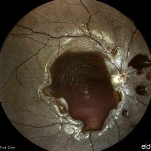
 CRVO
CRVO
Mar 29 2013 by Henry J. Kaplan, MD
Full blown ischemic CRVO with disc swelling, dilated and tortous veins, scattered hemorrhages and multiple cotton wool spots.
Condition/keywords: central retinal vein occlusion (CRVO), ischemic CRVO
-
 HIV Retinopathy
Aug 20 2014 by Andree Henaine-Berra, MD
Fundus photograph of the right eye of a HIV-positive male patient. The image shows multiple cotton wool spots and vascular tortuosity.
Photographer: Jorge Morales, MD. Hospital General "Dr. Manuel Gea Gonzalez". Mexico City
Condition/keywords: HIV retinopathy
-
 Acute Idiopathic Occlusive Retinal Vasculitis
May 31 2014 by Hamid Ahmadieh, MD
Color fundus photograph of the left eye of a 28-year-old woman with acute drop of vision due to occlusive retinal vasculitis leading to extensive nerve fiber layer infarction and retinal hemorrhages.
Photographer: Naghmeh Nozhat, Negah Eye Center, Tehran
Condition/keywords: color fundus photograph, cotton wool spots, retinal hemorrhage, retinal ischemia
-
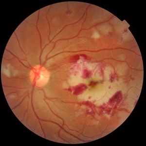
 Acute Necrotizing Retinal Vasculitis as Onset of Systemic Lupus Erythematosus.
Sep 3 2016 by ADRIANO FERREIRA
A 28-year-old white man was referred to the rheumatology clinic with gradually and rapid deterioration of the vision (both eyes). In this picture, we can observe cotton wool spots in the papillomacular area and extensive hemorrhages in posterior polo and in the middle periphery. Hard exudates are present in macular area (macular edema)
Photographer: Claudio Zett Lobo
Imaging device: TRC50DXi TOPCON
Condition/keywords: systemic lupus erythematosus (SLE) vasculitis, vasculitis
-
 Arterio-Venous Nipping, Venous Beading
Mar 1 2014 by Homayoun Tabandeh, MD, FASRS
Arterio-venous nipping, venous beading, and cotton wool spots in a patient with hypertensive and diabetic retinopathy.
Condition/keywords: arteriovenous nipping, cotton wool spots, venous beading
-
 Cotton Wool Spot
Jul 10 2013 by Jason S. Calhoun
Fundus photograph shows a young male with a single cotton wool spot just nasal to the macula in the right eye.
Photographer: Jason S. Calhoun, Department of Ophthalmology, Mayo Clinic Jacksonville, Florida
Condition/keywords: cotton wool spots, hypertension
-
 CRVO with Flame Hemorrhages
Oct 1 2012 by Jeffrey G. Gross, MD, FASRS
CRVO with flame hemorrhages and cotton wool spots 20/80.
Condition/keywords: 20/80, central retinal vein occlusion (CRVO), cotton wool spots
-
---thumb.jpg/image-square;max$300,300.ImageHandler) HIV Retinopathy
HIV Retinopathy
Feb 27 2013 by Henry J. Kaplan, MD
HIV retinopathy, left eye: multiple cotton wool spots. #2
Condition/keywords: cotton wool spots, HIV retinopathy
-
 Hypertensive Retinopathy
Jun 28 2013 by Jason S. Calhoun
Patient came in complaining of spots in vision in both eyes. VA was 20/25 - right eye and 20/20- left eye. Fundus exam reveals little hemorrhages with cotton wool spots due to hypertension and anemia.
Photographer: Jason S. Calhoun, Mayo Clinic Jacksonville, Florida
Imaging device: TOPCON TRC 50-EX
Condition/keywords: hypertensive retinopathy
-
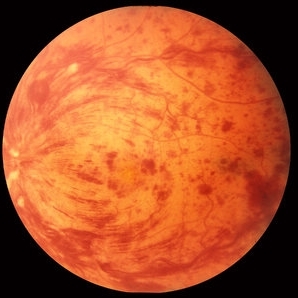
 SLE Retinopathy
Jul 10 2018 by Deepak Bhojwani, MS
Colour fundus montage image of a 33-year-old young lady with history of Systemic Lupus Erythematosus of 6 years showing classic SLE retinopathy with multiple cotton wool spots , few haemorrhages and multiple small vessel sheathing s/o SLE vasculitis.
Photographer: Deepak Bhojwani
Condition/keywords: systemic lupus erythematosus (SLE) retinopathy, systemic lupus erythematosus (SLE) vasculitis
-
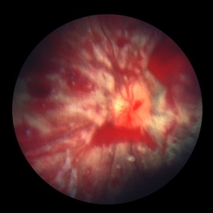
 Retinal Vasculitis with Hemorrhages and Cotton Wool Spots
Oct 16 2012 by Jeffrey G. Gross, MD, FASRS
Retinal vasculitis with hemorrhages and cotton wool spots.
Condition/keywords: cotton wool spots, retinal vasculitis
-
 Encephalitis with Retinal Cotton Wool Spots
Oct 15 2012 by Jeffrey G. Gross, MD, FASRS
Encephalitis with retinal cotton wool spots, right eye, 20/30.
Condition/keywords: cotton wool spots, encephalitis
-
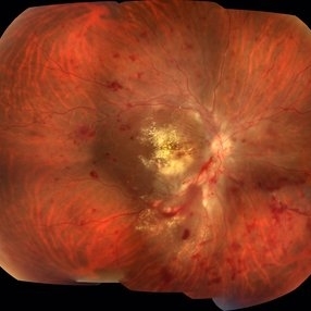
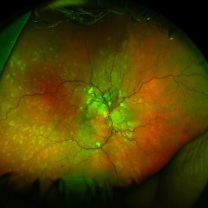
 Purtscher's Retinopathy with Diffuse Cotton Wool Spots CF
Oct 1 2012 by Jeffrey G. Gross, MD, FASRS
Purtscher's retinopathy with diffuse cotton wool spots CF.
Condition/keywords: cotton wool spots, Purtscher's retinopathy
-
---thumb.jpg/image-square;max$300,300.ImageHandler) HIV Retinopathy
HIV Retinopathy
Feb 27 2013 by Henry J. Kaplan, MD
HIV retinopathy, multiple cotton wool spots, right eye. #1
Condition/keywords: cotton wool spots, HIV retinopathy
-
 Anemic Retinopathy Related Retinal Hemorrhages
Nov 5 2019 by Chinmayi Vyas
Anemic retinopathy related retinal hemorrhages in a 24 years old male with Hb of 4.2gm/ dl. The manifestations of anemic retinopathy are nonspecific and may closely simulate hypertensive or diabetic retina. Retinal changes in anemia are cotton wool spots, venous tortuosity, and hemorrhages which may be present at all levels of the retina and choroid. All retinal hemorrhages can occur when Hb falls below 8 g/100 ml or if the platelet count falls below 50,000/cumm. The combination of severe anemia and thrombocytopenia is likely to produce retinal hemorrhages. The Roth’s spots or white centre hemorrhages are typically associated with bacterial endocarditis , anemia and other systemic conditions. The white center is suspected to represents focal ischemia, inflammatory or infectious infiltrate, fibrin or accumulation of neoplasticism cells.
Photographer: Dr Chinmayi Vyas
Condition/keywords: retinal hemorrhage
-
 Hypertensive Retinopathy, Right
Feb 23 2017 by Alla Goldberg, MD
Fundus photograph of 35-year-old man with severe hypertension (182/128).
Photographer: Sofia Rutiaga, UT Health McGovern Medical School, Cizik Eye Clinic
Condition/keywords: cotton wool spots, Elschnig's spots, hypertensive choroidopathy, hypertensive retinopathy, serous retinal detachment
-
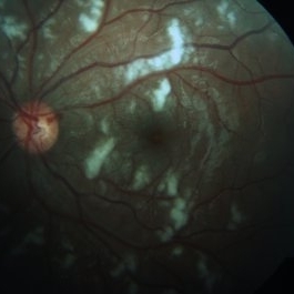 Purtscher's Retinopathy
Oct 16 2012 by S. Natarajan, MD, FASRS, FRCS (GLASGOW) , FICO, D.Sc, FELA
Fundus photograph of a 14-year-old male presenting with extensive cotton wool spots.
Photographer: Prof. Dr. S. Natarajan, Aditya Jyot Eye Hospital Pvt. Ltd., Mumbai, India
Condition/keywords: cotton wool spots, Purtscher's retinopathy
-
---thumb.jpg/image-square;max$300,300.ImageHandler) SLE Retinopathy
SLE Retinopathy
Feb 26 2013 by Henry J. Kaplan, MD
SLE retinopathy,l eft eye: multiple cotton wool spots and blot hemorrhages. #2
Condition/keywords: blot hemorrhages, cotton wool spots, systemic lupus erythematosus (SLE) retinopathy
-
 Shaken Baby Syndrome
Sep 20 2012 by Jeffrey G. Gross, MD, FASRS
Shaken Baby Syndrome with many hemorrhages and cotton wool spots
Condition/keywords: cotton wool spots, shaken baby syndrome
-
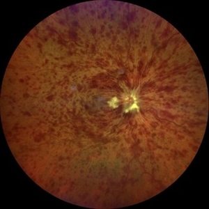
 Hypertensive retinopathy
Aug 24 2012 by Geoffrey G. Emerson, MD, PhD, FASRS
A 35-year-old man has headaches and decreased vision. The right eye measures 20/25 and the left eye measures 3/200. The blood pressure measures 180/110.
Photographer: Geoffrey Emerson, MD, PhD, Retina Center, Minneapolis
Condition/keywords: cotton wool spots, hypertensive retinopathy, papilledema
-
 Acute Necrotizing Retinal Vasculitis as Onset of Systemic Lupus Erythematosus.
Sep 3 2016 by ADRIANO FERREIRA
A 28-year-old white man was referred to the rheumatology clinic with gradually and rapid deterioration of the vision (both eyes). In this picture we can observe cotton wool spots in the papillomacular area and extensive hemorrhages in the left eye.
Photographer: Claudio Zett Lobo
Imaging device: TRC50DXi TOPCON
Condition/keywords: systemic lupus erythematosus (SLE) vasculitis, vasculitis
 Loading…