Search results (184 results)
-
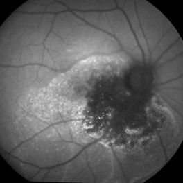
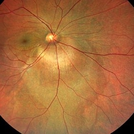 Circumscribed Choroidal Hemangioma with Serous Macular Retinal Detachment
Oct 2 2023 by Aditya S Kelkar, MS, FRCS, FASRS,FRCOphth
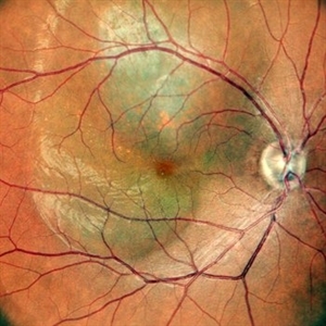
Fundus photograph of a 43-year-old male with a circumscribed choroidal hemangioma with serous macular retinal detachment associated with diminision of vision.
Photographer: Dr. Harsh Jain, National Institute of Ophthalmology
Imaging device: Clarus 500
Condition/keywords: choroidal hemangioma
-
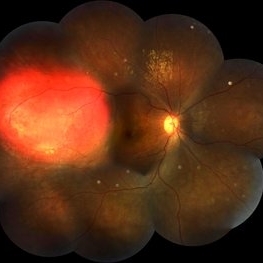 Focal Chroidal Hemangioma
Sep 18 2018 by Somnath Chakraborty, MD
Right eye fundus photo montage of a 17-year-old boy showing a focal choridal hemangioma temporally.
Photographer: Saptarshi Mehta, Retina Institute of Bengal
Condition/keywords: choroidal hemangioma
-
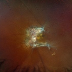
 Hemangioma of Retina
Mar 5 2025 by Virginia Gebhart
64 year old male with choroidal hemangioma in the macula and STA. Persistent IRF and new cuff of SRF compared to previous photos. BCVA CF@face. Pt has had PDT in the past with no significant improvement. Will observe closely
Photographer: Virginia Gebhart, Retina Consultants of Carolina
Imaging device: Optos California
Condition/keywords: hemangioma, inferior subretinal fluid
-
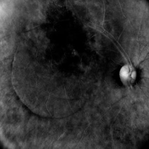
 Hemangioma of Retina (FAF)
Mar 5 2025 by Virginia Gebhart
Fundus autofluorescence of 64 year old male with choroidal hemangioma in the macula and STA. Persistent IRF and new cuff of SRF compared to previous photos. BCVA CF@face. Pt has had PDT in the past with no significant improvement. Will observe closely
Photographer: Virginia Gebhart, Retina Consultants of Carolina
Imaging device: Optos California
Condition/keywords: autofluorescence imaging, hemangioma, inferior subretinal fluid
-
 Indocyanine Green (ICG) of Circumscribed Choroidal Hemangioma (CCH)
Feb 6 2025 by Jack B Margines, MD, MHCI
Peripheral patchy hyperfluorescence is seen on this early image of ICG-A on a 53-year-old asymptomatic with an extramacular circumscribed choroidal hemangioma.
Photographer: W Ryan Miliam, CRA, OCT-C, University of California, Irvine Gavin Herbert Eye Institute
Imaging device: Optos
Condition/keywords: choroidal hemangioma, indocyanine green (ICG) angiography
-
 Sturge-Weber / Choroidal Hemangioma
Feb 27 2015 by David Callanan, MD
Male patient, Sturge-Weber / choroidal hemangioma.
Condition/keywords: choroidal hemangioma, Sturge-Weber syndrome
-
---thumb.jpg/image-square;max$300,300.ImageHandler) Sturge-Weber Diffuse Hemangioma and Retinal Detachment on B-scan
Sturge-Weber Diffuse Hemangioma and Retinal Detachment on B-scan
Apr 18 2014 by Susanna S. Park, MD, PhD
B-scan ultrasonogram of the right eye of an 8 year old Hispanic boy with Sturge -Weber Syndrome showing diffuse choroidal thickening from diffuse choroidal hemangioma and associated total exudative retinal detachment.
Photographer: Ellen Redenbo, University of California Davis Eye Center
Condition/keywords: B scan ultrasound, diffuse choroidal hemangioma, Sturge-Weber syndrome
-
 Sturge-Weber Syndrome
Sep 4 2024 by Virginia Gebhart
13 year old female with hemangioma of the retina and history of Sturge-Weber syndrome. Slightly larger compared to photos and ultrasound from 4 months ago. SRF inferior on ultrasound and clinical exam. Pt and mom will consider brachytherapy.
Photographer: Virginia Gebhart, Retina Consultants of Carolina
Imaging device: Optos California
Condition/keywords: diffuse choroidal hemangioma, Hemangioma, Sturge-Weber syndrome
-
 VHL Syndrome with Capillary Hemangioblastomas
Feb 26 2025 by Virginia Gebhart
39 year old female with choroidal hemangioma with capillary hemangioblastomas. Positive genetic testing for Von Hippel-Lindau Syndrome. Hemangioblastomas are stable compared to initial imaging in 2021. Pt started Welireg in Dec 2024, CNS tumors have started shrinking. No lesions in OD
Photographer: Virginia Gebhart, Retina Consultants of Carolina
Imaging device: Optos California
Condition/keywords: retinal capillary hemangioblastoma, Von Hippel-Lindau
-
 choroidal hemangioma
Feb 19 2018 by JEFFERSON R SOUSA, Tecg.º (Biomedical Systems Technology)
Patient female, 57 years old, with a low-vision complaint seven months ago. In specific ophthalmology exams, important changes were observed funducópicas suggestive of hemangioma de choroid.
Photographer: JEFFERSON R SOUSA - Study Center and Ophthalmological Research Dr. Andre M V Gomes, Institute Dr. Suel Abujamra São Paulo-Brazil
Condition/keywords: choroidal hemangioma
-
 Circumscribed Choroidal Hemangioma
Oct 20 2012 by Hyung-Woo Kwak, MD
Fundus and OCT examination showed an oval mass at the posterior pole with indistinct margins that blend with surrounding choroid. FA early phase showed hyperfluorescence.
-
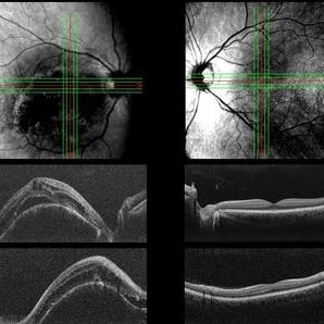 Both Eyes OCT in Case of Right Eye Choroidal Hemangioma
Nov 29 2024 by Anand Temkar
BE OCT of a 42 year old male, showing the elevation of the right eye retina along with the cystic spaces and subretinal fluid.
Photographer: Dr.Anand Temkar- Retina Foundation, Ahmedabad
Imaging device: Mirante
Condition/keywords: OCT
-
 Cavernous Choroidal Hemangioma
Jul 30 2013 by Jason S. Calhoun
Patient with cavernous choroidal hemangioma in the right eye. VA is 20/50 after manifest refraction. Patient also notices decreased vision with shadow close to her central vision in the right eye. Patient will undergo photodynamic therapy in the right eye.
Photographer: Jason S. Calhoun, Department of Ophthalmology, Mayo Clinic Jacksonville, Florida
Imaging device: TOPCON TRC 50-EX
Condition/keywords: cavernous choroidal hemangioma
-
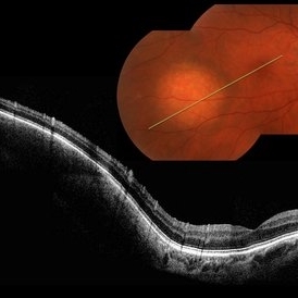
 CCH
CCH
Feb 6 2025 by Jack B Margines, MD, MHCI
SD-OCT of an extramacular Circumscribed Choroidal Hemangioma in an asymptomatic 53 year-old female
Photographer: Ryan Milam, University of California, Irvine Gavin Herbert Eye Institute
Imaging device: Zeiss Cirrus
Condition/keywords: Circumscribed Choroidal Hemangioma, OCT
-
 Central Macular Lesion, Choroidal Hemangioma, Staphyloma
Jul 11 2013 by Jerald A. Bovino, MD
No history.
Condition/keywords: central vascular lesion, staphyloma
-
 Choroidal Hemangioma
Jan 19 2021 by Stacie Neview
Ultra wide field fluorescein angiogram of a 44-year-old male with presumed central serous retinopathy. Based on extended ophthalmoscopy, diagnostic ocular ultrasonography, and retinal imaging, the choroidal tumor is most consistent with a choroidal hemangioma. A circumscribed choroidal hemangioma such as this one is unlikely to be associated with an underlying systemic condition and will be further monitored and assessed for possible treatment.
Photographer: Stacie Neview, COA, OSC
Imaging device: Optos California
Condition/keywords: choroidal hemangioma, early phase, fluorescein angiogram (FA), left eye, Optos, ultra-wide field imaging
-
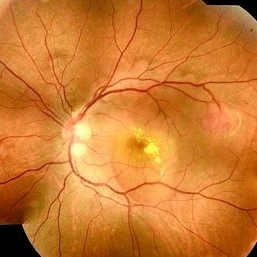 Choroidal Hemangioma
Mar 27 2021 by Paulo Horizonte
Fundus retinography of a 36-year-old man complaining of low progressive visual acuity in the left eye 2 years ago.
Photographer: Jeferson Sousa
Imaging device: Topcon
Condition/keywords: choroidal hemangioma
-
 CHOROIDAL HEMANGIOMA
Nov 21 2022 by Akansha Sharma
COLOU FUNDUS IMAGE OF A 32 YEAR OLD MALE WITH CHOROIDAL HEMANGIOMA WITH SUB RETINAL FLUID
Photographer: Dr. Akansha Sharma-Retina Foundation, Ahmedabad
Condition/keywords: choroidal hemangioma, subretinal fluid
-
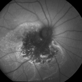
 CHOROIDAL HEMANGIOMA
Nov 21 2022 by Akansha Sharma
RETRO IMAGE OF A 32 YEAR OLD MALE WITH CHOROIDAL HEMANGIOMA WITH SUB RETINAL FLUID
Photographer: Dr. Akansha Sharma-Retina Foundation, Ahmedabad
Condition/keywords: choroidal hemangioma, subretinal fluid
-
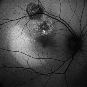 CHOROIDAL HEMANGIOMA
Nov 21 2022 by Akansha Sharma
BLUE-AUTOFLUORESCENCE IMAGE OF A 32 YEAR OLD MALE WITH CHOROIDAL HEMANGIOMA WITH SUB RETINAL FLUID
Photographer: Dr. Akansha Sharma-Retina Foundation, Ahmedabad
Condition/keywords: choroidal hemangioma, subretinal fluid
-
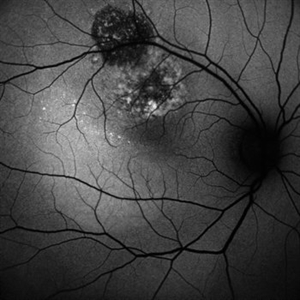 CHOROIDAL HEMANGIOMA
Nov 21 2022 by Akansha Sharma
GREEN-AUTOFLUORESCENCE IMAGE OF A 32 YEAR OLD MALE WITH CHOROIDAL HEMANGIOMA WITH SUB RETINAL FLUID
Photographer: Dr. Akansha Sharma-Retina Foundation, Ahmedabad
Condition/keywords: choroidal hemangioma, subretinal fluid
-
 Choroidal Hemangioma
Jul 30 2024 by Korey Starkey
ICG image of a77 year-old female with choroidal hemangioma. The physician states the hypercyanesence in the right eye is consistent with hemangioma but no typical late washout observed. He also notes high internal reflectivity make hemangioma possible. Patients vision at time of imaging VA OD: sc20/200 PH20/60-1; plan to follow patient at 6 month intervals at this time.
Photographer: Korey Starkey
Imaging device: Optos
Condition/keywords: Choroidal Hemangioma, Fluorescein angiography, indocyanine green (ICG) angiography, Optos
 Loading…