Search results (632 results)
-
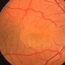
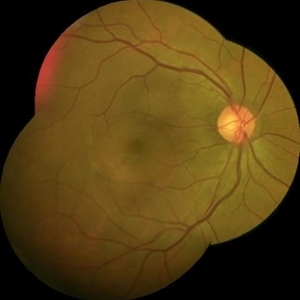
 Central Serous Retinopathy
Mar 19 2024 by Corey Grant
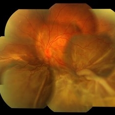
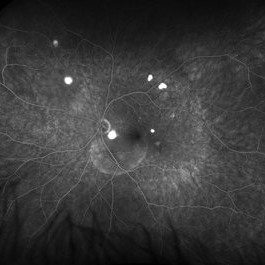
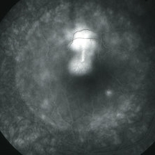
Ultra Wide-Field Fundus Autofluorescence Imaging of a 37 year old female with Central Serous Retinopathy affecting her right eye. Patient Visual Acuity was 20/20 in both eyes. Patient reported black spots in her vision onset three years ago, with associating flashes of light. Patient reports history of cortisone back injections a few years ago and denies Flonase use. The physician stated that there is hyperautofluorescence in the area of gutter of Sub-Retinal Fluid which likely happened from CSR.
Photographer: Corey Grant, OSC
Imaging device: OPTOS CALIFORNIA RGB
Condition/keywords: Central Serous Chorioretinopathy (CSR), central serous retinopathy (CSR), fundus autofluorescence (FAF), Guttering, hyperautofluorescence, inferior retina, OPTOS, Retina, Right Eye, subretinal fluid, ULTRA WIDE FIELD
-
 Multifocal CSR With Exudative RD
May 19 2017 by Manish Nagpal, MD, FRCS (UK), FASRS
A 30-year-old male diagnosed elsewhere as VKH was started on heavy steroids and he developed multiple serous elevations and OS developed a exudative RD.
Photographer: POOJA BAROT
Condition/keywords: central serous retinopathy (CSR), multifocal central serous chorioretinopathy (CSCR), Vogt-Koyanagi-Harada
-
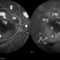
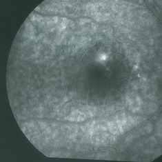
 Central Serous Chorioretinopathy
Jan 25 2022 by Olivia Rainey
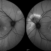
Widefield fundus autofluorescence of a 60-year-old male with Central Serous Chorioretinopathy affecting both eyes. Chronic history of CSR followed with observation without treatment prior to presenting at our office. The physician noted significant findings on exam and imaging with multifocal areas of inactive and active changes in the right eye and subfoveal subretinal fluid with recent visual decline in the left eye. There are hyper and hypoautofluorescent changes, consistent with CSR.
Photographer: Olivia Rainey, OCT-C, COA
Imaging device: Heidelberg Spectralis
Condition/keywords: 55-degrees, central serous chorioretinopathy (CSCR), central serous retinopathy (CSR), chronic central serous chorioretinopathy (CSCR), fundus autofluorescence (FAF), heidelberg spectralis, left eye
-
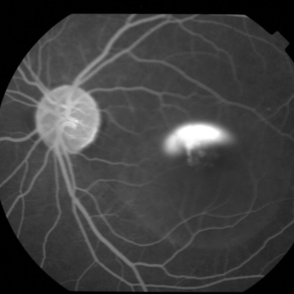
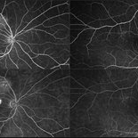
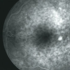
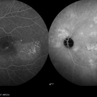 Central Serous Chorioretinopathy
Jan 25 2022 by Olivia Rainey
Late phase widefield fluorescein angiography of a 60-year-old male with Central Serous Chorioretinopathy. Chronic history of CSR followed with observation without treatment prior to presenting at our office. The physician noted subfoveal subretinal fluid with recent visual decline. FA shows multifocal leakage and ICG shows hypercyanescence. OCTA, ICG, and FA consistent with CSR, and without concern for CNVM thus will observe without anti-VEGF at this time. PDT therapy recommended.
Photographer: Olivia Rainey, OCT-C, COA
Imaging device: Heidelberg Spectralis
Condition/keywords: 55-degrees, central serous chorioretinopathy (CSCR), central serous retinopathy (CSR), chronic central serous chorioretinopathy (CSCR), fluorescein angiogram (FA), heidelberg spectralis, indocyanine green (ICG) angiography, left eye
-
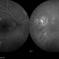 Central Serous Chorioretinopathy
Jan 25 2022 by Olivia Rainey
Late phase widefield fluorescein angiography of a 60-year-old male with Central Serous Chorioretinopathy. Chronic history of CSR followed with observation without treatment prior to presenting at our office. The physician noted significant findings on exam and imaging with multifocal areas of inactive and active changes OD. FA shows superotemporal macular leakage, subtle inferonasal macular leakage and staining as well as multifocal hypercyanescence on ICG. Fortunately foveal sparing and thus observation is recommended at this time OD.
Photographer: Olivia Rainey, OCT-C, COA
Imaging device: Heidelberg Spectralis
Condition/keywords: 55-degrees, central serous chorioretinopathy (CSCR), central serous retinopathy (CSR), chronic central serous chorioretinopathy (CSCR), fluorescein angiogram (FA), fluorescein leakage, heidelberg spectralis, indocyanine green (ICG) angiography, late phase
-
 Central Serous Choroidopathy - Angiography
Jun 27 2018 by Gabriel Costa Andrade, PhD
46-year-old male with central serous choroidopathy in left eye. VA OS cc 20/40. Late phase FA photo shows multiple foci of leakage.
Photographer: Gabriel Andrade, RETINA CLINIC, São Paulo, BRAZIL
Imaging device: Optos California
Condition/keywords: central serous retinopathy (CSR)
-
 Central Serous Choroidopathy, CSR, with Foci of Leakage
Oct 9 2012 by James B. Soque, CRA, OCT-C, COA, FOPS
50 y/o WM with Central Serous Choroidopathy Left eye. VA OS cc 20/80. Topcon 3D 1000 SD OCT image reveals Sub RPE detachment in several locations, and subretinal fluid blister. Color, Early, and Late phase FA photos enclosed. FA shows obvious ‘smoke stack’ appearance of leakage in superonasal fovea, and 3 other foci of leakage. Late FA Photo shown.
Photographer: James Soque CRA COA
Imaging device: Topcon TRC 50 EX, with OIS V 10.5.74 Software. 5 MP Camera
Condition/keywords: central serous chorioretinopathy (CSCR), central serous retinopathy (CSR)
-
 Central Serous Retinopathy with Smokestack Pattern
Aug 15 2015 by Thomas A. Ciulla, MD, MBA, FASRS
Central serous retinopathy with smokestack pattern.
Photographer: Charlotte Harris
Condition/keywords: central serous retinopathy (CSR)
-
 Multifocal CSR OD
May 19 2017 by Manish Nagpal, MD, FRCS (UK), FASRS
A 30-year-old male diagnosed elsewhere as VKH was started on heavy steroids and he developed multiple serous elevations and OS developed a exudative RD.
Photographer: POOJA BAROT
Condition/keywords: central serous retinopathy (CSR), multifocal central serous chorioretinopathy (CSCR), Vogt-Koyanagi-Harada
-
 Multifocal CSR OS FA and ICG
May 19 2017 by Manish Nagpal, MD, FRCS (UK), FASRS
A 30-year-old male diagnosed elsewhere as VKH was started on heavy steroids and he developed multiple serous elevations and OS developed a exudative RD.
Photographer: pooja barot
Condition/keywords: central serous retinopathy (CSR), Vogt-Koyanagi-Harada
-
 Multifocal CSR FA & ICG
May 19 2017 by Manish Nagpal, MD, FRCS (UK), FASRS
A 30-year-old male diagnosed elsewhere as VKH was started on heavy steroids and he developed multiple serous elevations and OS developed a exudative RD. We immediately asked the patient to stop steroids and when he followed up after a month lesions had flattened and he had recovered to 20/40 in both eyes.. he is still undergoing further follow up at this stage...
Photographer: pooja barot
Imaging device: heidelberg
Condition/keywords: central serous retinopathy (CSR), multifocal central serous chorioretinopathy (CSCR), Vogt-Koyanagi-Harada
-
 Central Serous Retinopathy - Early FA
Feb 22 2016 by Ahmad B. Tarabishy, MD
48-year-old male with newly diagnosed central serous retinopathy OD.
Photographer: Jessica Armbruster
Imaging device: Topcon TRC-50EX
Condition/keywords: central serous retinopathy (CSR)
-
 Central Serous Retinopathy with Smokestack
Nov 5 2019 by Nichole Lewis
40-year-old male with central serous retinopathy with smokestack.
Photographer: Nichole Lewis
Imaging device: Optos
Condition/keywords: central serous retinopathy (CSR), smokestack
-
 Optic Disc Pit
Jun 4 2014 by Henry J. Kaplan, MD
Optic disc pit in the temporal part of optic nerve with associated CSR.
Condition/keywords: central serous retinopathy (CSR), optic disc pit
-
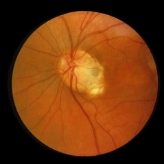
 Chronic Central Serous Chorioretinopathy
Mar 29 2019 by Nichole Lewis
54-year-old male with chronic central serous retinopathy with focal sub-retinal fluid and widespread retinal pigment epithelium changes. History of micropulse laser. Patient is HLA-B27 positive with quiescent iritis. VA 20/20.
Photographer: Nichole Lewis
Imaging device: Optos
Condition/keywords: central serous retinopathy (CSR), retinal pigment epithelium (RPE) changes, subretinal fluid
-
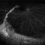
 Bilateral Central Serous Retinopathy
Mar 26 2019 by Gary R. Cook, MD, FACS
Late-phase frame of FA of 37-year-old white male with acute CSR OD showing pooling of dye beneath the small central RPED centrally, a smokestack-type leak from the RPE defect just above it, and mild late pooling of dye outlining the large neurosensory macular detachment; VA = 20/80-1.
Imaging device: Topcon VT-50
Condition/keywords: central serous retinopathy (CSR), FA late phase, FA late phase leakage, neurosensory detachment of retina
-
 Bilateral Central Serous Retinopathy
Mar 26 2019 by Gary R. Cook, MD, FACS
Right eye of a 37-year-old white male with a history of bilateral CSR showing a 2 DD NSRD centrally in his symptomatic OD; VA = 20/20-2.
Imaging device: Topcon VT-50
Condition/keywords: central serous retinopathy (CSR), neurosensory detachment of retina
-
 Bilateral Central Serous Retinopathy
Mar 26 2019 by Gary R. Cook, MD, FACS
Asymptomatic left eye of a 37-year-old white male with a history of previous CSR OS showing some focal RPE depigmentation perifoveally and subretinic deposits temporally; no NSRD is present; VA = 20/15+3.
Imaging device: Topcon VT-50
Condition/keywords: central serous retinopathy (CSR), resolved subretinal fluid, retinal pigment epithelium (RPE) changes
-
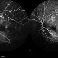
 Bilateral Central Serous Retinopathy
Mar 26 2019 by Gary R. Cook, MD, FACS
Late-phase fluorescein angiogram image of the right eye of a 37-year-old white male showing pinpoint leak with late diffusion of dye from it superiorly and RPE irregularities nasal to fovea in a case of bilateral central serous retinopathy; VA = 20/20-2.
Imaging device: Topcon VT-50
Condition/keywords: central serous retinopathy (CSR), FA late phase, fluorescein angiogram (FA)
-
 Bilateral Central Serous Retinopathy
Mar 26 2019 by Gary R. Cook, MD, FACS
Mid-phase fluorescein angiogram frame of a pinpoint leak in the temporal macula OS of a 37-year-old white male with bilateral central serous retinopathy; VA = 20/15+3.
Imaging device: Topcon VT-50
Condition/keywords: central serous retinopathy (CSR), FA mid phase, fluorescein angiogram (FA)
-
 Central Serous Chorioretinopathy
Mar 3 2020 by Sham Talati, DOMS
A patient who had CSR in RE.
Photographer: Dr. Ashok Talati, Dr.Talati's Eye Hospital, Ahmedabad
Condition/keywords: central serous chorioretinopathy (CSCR), central serous retinopathy (CSR)
-
 Central serous chorioretinopathy
Apr 30 2015 by Mariam A Al-Feky, MD
A case of CSR phtographed on the Heidelberg fundus camera with multicolor image, infrared, blue reflectance and green reflectence predye injection, postdye injection and during dye injection and last image for the OCT. Fundus examination after dye injection showed a green spot nasal that was not detected predye injection. Multicolor image was retaken and that green spot is well evident in the multicolor image, the infrared relectance, blue and green reflectance. That green spot is corresponding to the leaky point in FFA and to a PED in OCT.
Photographer: Mariam AL-Feky
Condition/keywords: central serous retinopathy (CSR), leakage
-
 Central Serous Chorioretinopathy (CSC)
Oct 16 2012 by S. Natarajan, MD, FASRS, FRCS (GLASGOW) , FICO, D.Sc, FELA
Middle-aged male came with small PED 4 months back; now this has progressed to a larger PED with SRF underneath the fovea.
Photographer: Prof. Dr. S. Natarajan
Condition/keywords: central serous chorioretinopathy (CSCR), central serous retinopathy (CSR), pigment epithelial detachment (PED), subretinal fibrosis
-
 Central Serous Chorioretinopathy Associated With Steroids
Jul 31 2016 by Mitzy E Torres Soriano, MD
FA (late phase) of bilateral central serous chorioretinopathy associated with steroids.
Photographer: Mitzy E. Torres Soriano. Centro de la Visión Gordon-Manavella. Rosario, Argentina
Imaging device: TOPCON
Condition/keywords: bilateral chronic central serous retinopathy, central serous retinopathy (CSR)
-
 Central Serous Chorioretinopathy Treated with PDT
Oct 17 2017 by Theodore Leng, MD, MS, FASRS
Central serous chorioretinopathy 1 month after treatment with PDT.
Condition/keywords: central serous retinopathy (CSR), idiopathic central serous choroidopathy (ICSC), photodynamic therapy
 Loading…