Search results (261 results)
-
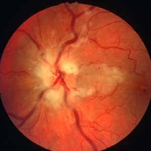
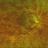
 Venous Beading
Nov 4 2021 by Stefanie Palmer
Venous Beading in a patient with both PDR and CRVO.
Photographer: Stefanie Palmer, CRA
Imaging device: Topcon
Condition/keywords: central retinal vein occlusion (CRVO), diabetic retinopathy, proliferative diabetic retinopathy (PDR), venous beading
-
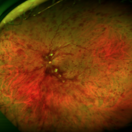
 Bilateral CRVO and PDR
Nov 4 2021 by Stefanie Palmer
Patient with both PDR and CRVO, 34 year old female, post-COVID.
Photographer: Stefanie Palmer, CRA
Imaging device: Topcon
Condition/keywords: central retinal vein occlusion (CRVO), COVID-19, diabetic retinopathy, proliferative diabetic retinopathy (PDR), venous beading
-
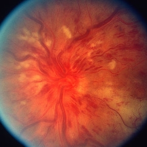
---thumb.jpg/image-square;max$300,300.ImageHandler) Central Retinal Vein Occlusion
Central Retinal Vein Occlusion
Oct 30 2012 by Lihteh Wu, MD
35-year-old hypertensive man with an acute CRVO. Notice the peripapillary cotton wool spots, superficial flame shaped hemorrhages and deeper dot and blot hemorrhages in all 4 quadrants. This is the typical blood and thunder appearance of a CRVO.
Condition/keywords: central retinal vein occlusion (CRVO), cotton wool spots
-
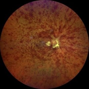
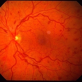
 CRVO
CRVO
Apr 22 2017 by Gabriel Costa Andrade, PhD
Panoramic retinography (Optos® California) of the right eye of a 48-year-old female patient with a history of low-vision in the right eye 2 months ago. At the exam presented visual acuity of 20/200 in the right eye and 20/20 in the left eye. Angiography shows diffuse perivascular leakage associated with areas of hypoperfusion in macula and periphery.
Photographer: Gabriel Andrade
Imaging device: Optos® California
Condition/keywords: central retinal vein occlusion (CRVO)
-
 Bilateral CRVO and PDR
Nov 4 2021 by Stefanie Palmer
Patient with both PDR and CRVO, 34 year old female, post-COVID.
Photographer: Stefanie Palmer, CRA
Imaging device: Topcon
Condition/keywords: central retinal vein occlusion (CRVO), COVID-19, diabetic retinopathy, proliferative diabetic retinopathy (PDR), venous beading
-
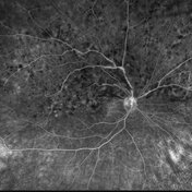
 Central Retinal Vein Occlusion
Jan 21 2022 by Olivia Rainey
Ultra-widefield fluorescein angiogram of a 23-year-old female with a Central Retinal Vein Occlusion affecting her left eye. The patient presented on 12/22/2021 cc20/40-2 vision in the left eye. The patient reported recent trauma of being hit with a fist on both sides of face followed by vision loss. The patient has history of Hashimoto's thyroid disease. The following labs have been ordered, PT, PTT, CBC, antithrombin III activity, protein C, protein S, Factor V Leiden mutation, Prothrombin (G20210A), lipid panel, HbA1c, quantiferon gold, RPR, and CXR.
Photographer: Olivia Rainey, OCT-C, COA
Imaging device: Optos California
Condition/keywords: central retinal vein occlusion (CRVO), disc leakage, fluorescein angiogram (FA), fluorescein leakage, left eye, non-ischemic central retinal vein occlusion (CRVO), Optos, trauma, ultra-wide field imaging
-
---thumb.jpg/image-square;max$300,300.ImageHandler) Central Retinal Vein Occlusion
Central Retinal Vein Occlusion
Oct 30 2012 by Lihteh Wu, MD
35-year-old hypertensive man with an acute CRVO. Notice the peripapillary cotton wool spots, superficial flame shaped hemorrhages and deeper dot and blot hemorrhages in all 4 quadrants. This is the typical blood and thunder appearance of a CRVO.
Condition/keywords: central retinal vein occlusion (CRVO), cotton wool spots
-
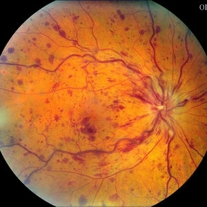
 Central Retinal Vein Occlusion associated with disc edema
Oct 19 2023 by Gabriel Costa Andrade, PhD
53-year-old woman with an acute CRVO. The patient has a history of breast cancer undergoing treatment with systemic chemotherapy. Notice the peripapillary cotton wool spots, superficial flame shaped hemorrhages and deeper dot and blot hemorrhages in all 4 quadrants.
Photographer: Gabriel Andrade
Condition/keywords: central retinal vein occlusion (CRVO), macular edema, Retina
-
 CRVO
CRVO
Mar 29 2013 by Henry J. Kaplan, MD
Full blown ischemic CRVO with disc swelling, dilated and tortous veins, scattered hemorrhages and multiple cotton wool spots.
Condition/keywords: central retinal vein occlusion (CRVO), ischemic CRVO
-
 CRVO with cilioretinal artery occlusion
Jan 11 2013 by Alex P. Hunyor, MD
Nonischaemic central retinal vein obstruction (CRVO) with cilioretinal artery occlusion.
Condition/keywords: central retinal vein occlusion (CRVO), cilioretinal artery occlusion
-
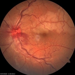
 CRVO with papillophlebitis
Oct 22 2020 by Gabriel Costa Andrade, PhD
Fundus photograph of an 43-year-old man with CRVO and papillophlebitis.
Photographer: Gabriel Andrade
Condition/keywords: central retinal vein occlusion (CRVO)
-
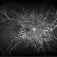
 HRVOFA
Aug 13 2021 by Jeffrey Barker
Hemi-retinal vein occlusion. Right-eye.
Photographer: Jeffrey P. Barker, B.S. Retina Vitreous Surgeons of C.N.Y.
Condition/keywords: central retinal vein occlusion (CRVO), hemi CRVO
-
 Non Ischemic CRVO
Mar 29 2013 by Henry J. Kaplan, MD
Non ischemic CRVO.
Condition/keywords: central retinal vein occlusion (CRVO)
-
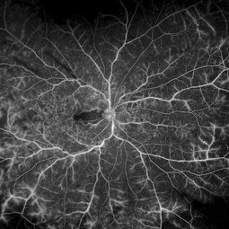
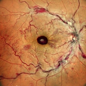
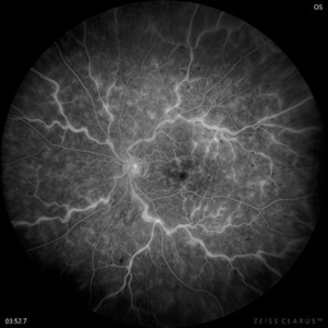 Retinal Lightning
Apr 24 2022 by Mariam Cernichiaro-Espinosa, MD
Late phase venous leakage on IV fluorescein angiography from a 32-year-old male with central retinal vein occlusion (CRVO).
Photographer: Mariam Cernichiaro-Espinosa, Asociación para Evitar la Ceguera, I.A.P. Mexico City, Mexico.
Imaging device: Zeiss Clarus
Condition/keywords: central retinal vein occlusion (CRVO)
-
 Central Retinal Vein Occlusion
Feb 28 2021 by AGNES KIM
Fundus photograph of unilateral CRVO.
Photographer: Agnes Kim
Condition/keywords: central retinal vein occlusion (CRVO)
-
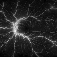
 Central Retinal Vein Occlusion
Sep 27 2024 by Korey Starkey
Fluorescein angiogram of a 75 year old patient with central retinal vein occlusion. FA shows areas of patchy ischemia and petaloid leakage. Patient is being treated with anti-vegf treatments at this time.
Photographer: Korey Starkey
Condition/keywords: central retinal vein occlusion (CRVO), FLUORESCEIN ANGIOGRAPHY, ischemia, macular edema, petaloid leakage, ultra-widefield image
-
 Central Retinal Vein Occlusion
Mar 27 2014 by Jason S. Calhoun
Patient in with mild blurred vision in the right eye. Fundus exam shows CRVO with scattered retinal hemorrhages in the right eye.
Photographer: Jason S. Calhoun, Mayo Clinic Jacksonville, Department of Ophthalmology
Imaging device: TOPCON TRC 50-EX
Condition/keywords: central retinal vein occlusion (CRVO)
-
 Central Retinal Vein Occlusion with Cilioretinal Artery Occlusion
Oct 21 2020 by Rutul R Patel, MD Ophthalmology
Fundus photograph of left eye of 37-year-old female who presented with sudden painless loss of vision in left eye due to CRVOwith CLRAO.
Photographer: Vidhi Bavishi, Shivjyoti Eye Hospital
Imaging device: TOPCON MAESTRO
Condition/keywords: central retinal vein occlusion (CRVO), cilioretinal artery occlusion
-
 Central Retinal Vein Occlusion with Foveal Hemorrhage
Apr 17 2025 by Malvika Singh
Fundus photograph of a 41 year-old, male, with a central retinal vein occlusion and a foveal sub-internal limiting membrane hemorrhage.
Photographer: Dr Malvika Singh, Retina Foundation, Ahmedabad, India
Imaging device: Mirante SLO/OCT
Condition/keywords: central retinal vein occlusion (CRVO), macular hemorrhage
-
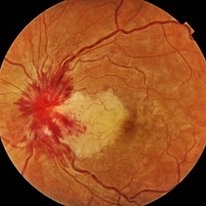
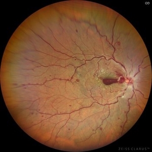 Central Retinal Vein Occlusion with Preretinal Hemorrhage
Mar 16 2021 by MOHIT GUPTA
Fundus photograph of right eye of a young male after 2nd dose of Covishield vaccine presented to us with central retinal vein occlusion and preretinal hemorrhage at macula in right eye.
Photographer: Dr Mohit Gupta , Prakash Netra Kendr, Lucknow, India
Imaging device: zeiss clarus
Condition/keywords: central retinal vein occlusion (CRVO), preretinal hemorrhage
-
 Central Retinal Vein Occlusion with Retinal Neovascularization
Jan 19 2022 by Olivia Rainey
Ultra-widefield fluorescein angiogram of a 56-year-old male with a Central Retinal Vein Occlusion with Retinal Neovascularization affecting his left eye. The patient presented on 1/19/2022 with scNLP vision in the left eye. The patient has good PRP, however areas of ischemia still remain untreated by laser. He also has severe neovascular glaucoma contributing to his poor vision.
Photographer: Olivia Rainey, OCT-C, COA
Imaging device: Optos California
Condition/keywords: central retinal vein occlusion (CRVO), FA early phase, fluorescein angiogram (FA), hemorrhage, ischemic CRVO, left eye, neovascular glaucoma, Optos, pan-retinal photocoagulation (PRP), retinal ischemia, retinal neovascularization, ultra-wide field imaging
-
 Central Retinal Vein Occlusion with Severe Retinal Ischemia
Jan 19 2022 by Olivia Rainey
Ultra-widefield fluorescein angiogram of a 56-year-old male with a Central Retinal Vein Occlusion with Severe Retinal Ischemia affecting his right eye. The patient presented on 1/19/2022, sc20/20-2 vision in the right eye. The patient has had a good response to Eylea with complete resolution of edema. The physician is considering PRP to ischemic periphery in the future and given the degree of ischemia in both eyes, she recommends that the patient's PCP check carotid Doppler US.
Photographer: Olivia Rainey, OCT-C, COA
Imaging device: Optos California
Condition/keywords: central retinal vein occlusion (CRVO), FA late phase, fluorescein angiogram (FA), ischemic CRVO, Optos, retinal ischemia, ultra-wide field imaging
-
---thumb.jpg/image-square;max$300,300.ImageHandler) Cilioretinal Artery Occlusion with Central Retinal Vein Occlusion
Cilioretinal Artery Occlusion with Central Retinal Vein Occlusion
Mar 9 2013 by Gabriela Lopezcarasa Hernandez, MD
A 46-year-old male with decrease in visual acuity in left eye and central scotoma.
Photographer: Araceli Rojas Arriaga, Hospital Angeles Lomas, Mexico
Imaging device: Zeiss FF4
Condition/keywords: central retinal vein occlusion (CRVO), cilioretinal artery occlusion
-
 Combined central retinal vein occlusion and branch retinal arteriolar occlusion
Sep 13 2022 by Ruchir Mehta, DO, DNB, FRCS
Fundus photograph of left eye of a 63 years old female with known type 2 DM and HTN showing combined central retinal venous occlusion and superior branch retinal arteriolar occlusion
Photographer: Ruchir Mehta, Mehta Superspeciality Eye Hospital, Jamnagar, Gujarat, India
Imaging device: Zeiss Visucam 500
Condition/keywords: branch retinal artery occlusion (BRAO), central retinal vein occlusion (CRVO), COMBINED
-
 CRVO
CRVO
Apr 15 2021 by David L Kilpatrick, MD
A CRVO in a 72-year-old female with a history of hypertension.
Photographer: Kyle McClellan
Imaging device: Optos
Condition/keywords: central retinal vein occlusion (CRVO)
 Loading…