Search results (162 results)
-
 Branch Retinal Artery Occlusion With Calcium Embolus at the Disc - Fundus Autofluorescence Imaging (FAF)
Apr 7 2018 by Rameez N Hussain, MD
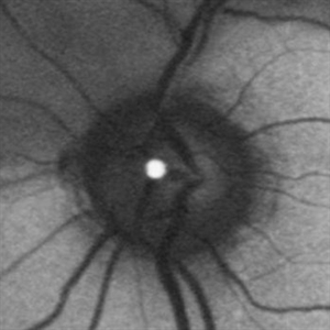
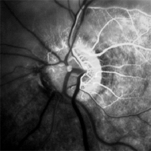
Acute branch retinal artery occlusion with a calcium embolus at the disc which is hyper autofluorescent in fundus autofluorescence imaging (FAF) -resembles an LED light source ('LED sign').
Photographer: DR RAMEEZ N HUSSAIN
Imaging device: Heidelberg Spectralis
Condition/keywords: branch retinal artery occlusion (BRAO), embolus, fundus autofluorescence (FAF), retinal edema
-
 BRAO Rianto AF
Apr 12 2014 by Sjakon G Tahija, MD
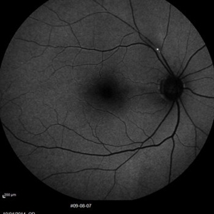
Auto fluorescence fundus image of a 70-year-old man with a superior temporal branch retinal artery occlusion. The emboli can be very clearly seen as the white dot of AF blockage.
Photographer: Avris Siahaan
Imaging device: Heidelberg Spectralis
Condition/keywords: branch retinal artery occlusion (BRAO)
-
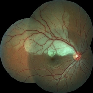
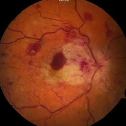
 Branch Retinal Artery Occlusion With Calcium Embolus at the Disc - Fundus Photo
Apr 7 2018 by Rameez N Hussain, MD
Acute branch retinal artery occlusion with a calcium embolus at the disc with retinal whitening in the area of retinal edema.
Photographer: DR RAMEEZ N HUSSAIN
Imaging device: zeiss
Condition/keywords: branch retinal artery occlusion (BRAO), embolus, fundus photograph, retinal edema
-
 Branch Retinal Artery Occlusion With Calcium Embolus at the Disc - Fundus Autofluorescence Imaging (FAF)
Apr 7 2018 by Rameez N Hussain, MD
Acute branch retinal artery occlusion with a calcium embolus at the disc which is hyper autofluorescent in fundus autofluorescence Imaging (FAF) -resembles an LED light source ('LED sign').
Photographer: DR RAMEEZ N HUSSAIN
Imaging device: Heidelberg Spectralis
Condition/keywords: branch retinal artery occlusion (BRAO), embolus, fundus autofluorescence (FAF), retinal edema
-
 Branch Retinal Artery Occlusion With Calcium Embolus at the Disc - Fundus Fluorescence Angiogram (FA)
Apr 7 2018 by Rameez N Hussain, MD
Acute branch retinal artery occlusion with a calcium embolus at the disc which is hyperfluorescent in FA.
Photographer: DR RAMEEZ N HUSSAIN
Imaging device: Zeiss
Condition/keywords: branch retinal artery occlusion (BRAO), embolus, fluorescein angiogram (FA), retinal edema
-
 Branch Retinal Artery Occlusion
Sep 9 2018 by Gabriela Lopezcarasa Hernandez, MD
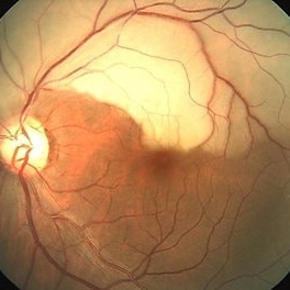
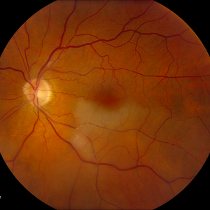
88-year-old female patient with sudden decrease in visual acuity and scotoma in left eye, please notice the widening retina due to retinal edema of branch occlusion with hollenhorst plaque in the artery and the optic nerve.
Photographer: Araceli Rojas
Imaging device: Zeiss FF4
Condition/keywords: branch retinal artery occlusion (BRAO)
-
 Branch Retinal Artery Occlusion
Sep 9 2018 by Gabriela Lopezcarasa Hernandez, MD
88-year-old female patient with sudden decrease in visual acuity and scotoma in left eye, please notice the branch occlusion with hollenhorst plaque and the delay perfusion in the involved arteria.
Photographer: Araceli Rojas
Imaging device: Zeiss FF4
Condition/keywords: branch retinal artery occlusion (BRAO)
-
 Branch Retinal Artery Occlusion
Mar 27 2018 by Nichole Lewis
Branch retinal artery occlusion with a Hollenhorst Plaque.
Photographer: Nichole Lewis
Condition/keywords: branch retinal artery occlusion (BRAO), hollenhorst plaque
-
 Branch Retinal Artery Occlusion
Dec 2 2019 by Kristen Wagner
Fundus Photo of a left eye with a branch retinal artery occlusion. Vision was DVA cc 20/40.
Photographer: Kristen Wagner, COT, OSC, Ophthalmic Photographer, Tennessee Retina
Condition/keywords: branch retinal artery occlusion (BRAO)
-
 BRAO
BRAO
Jun 29 2014 by John S. King, MD
BRAO.
Photographer: Wayne A Ladlee Jr
Condition/keywords: branch retinal artery occlusion (BRAO), embolic
-
 BRAO d/t cat scratch disease - FA 00:18 min.
Jan 2 2013 by Roy Schwartz, MD
A 38-year-old male complained of a grey spot in visual field in his left eye. On clinical exam BRAO in LE, confirmed by FA, as seen in picture. Image shows delayed filling of artery. Serology for bartonella was positive.
Photographer: Galit Yair-Pur
Condition/keywords: branch retinal artery occlusion (BRAO), cat scratch retinitis
-
 Combined central retinal vein occlusion and branch retinal arteriolar occlusion
Sep 13 2022 by Ruchir Mehta, DO, DNB, FRCS
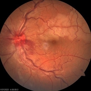
Fundus photograph of left eye of a 63 years old female with known type 2 DM and HTN showing combined central retinal venous occlusion and superior branch retinal arteriolar occlusion
Photographer: Ruchir Mehta, Mehta Superspeciality Eye Hospital, Jamnagar, Gujarat, India
Imaging device: Zeiss Visucam 500
Condition/keywords: branch retinal artery occlusion (BRAO), central retinal vein occlusion (CRVO), COMBINED
-
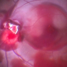
 IOFB Over Disc With Blood Stained Hyaloid and BRAO
Feb 25 2017 by Manish Nagpal, MD, FRCS (UK), FASRS
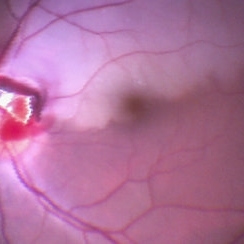
Intraoperative photo of a foreign body piercing the inferotemporal margin of disc revealing a infero temporal BRAO immediately after hyaloid removal.
Photographer: MANISH NAGPAL
Imaging device: Still captured from a 3 chip HD camera attached to a microscope
Condition/keywords: branch retinal artery occlusion (BRAO), intraocular foreign body
-
 IOFB Over Disc With Blood Stained Hyaloid and BRAO
Feb 25 2017 by Manish Nagpal, MD, FRCS (UK), FASRS
Intraoperative photo of a foreign body piercing the inferotemporal margin of disc revealing a infero temporal BRAO and blood stained hyaloid just prior to removal of hyaloid and IOFB.
Photographer: manish nagpal
Imaging device: Still captured from 3 Chip HD camera on microscope
Condition/keywords: branch retinal artery occlusion (BRAO), intraocular foreign body
-
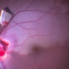 IOFB with BRAO
Feb 9 2017 by Manish Nagpal, MD, FRCS (UK), FASRS
Intraoperative photo of a IOFB impacting on the inferotemporal margin of disc leading to a BRAO. This picture is taken moments after dissecting the IOFB from the impact site and bringing it over the retinal surface for eventual removal.
Photographer: Manish Nagpal
Imaging device: Still captured from a 3 chip HD camera on microscope
Condition/keywords: branch retinal artery occlusion (BRAO), intraocular foreign body
-
 Superior Hemi-Central Retinal Artery Occlusion
Apr 24 2024 by Mosab Salah
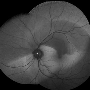
Fundus photograph -inverted view- taken by smartphone fundus photography, of a young man with sudden onset altitudinal field defect, a Superior Hemi-Central Retinal Artery Occlusion noted.
Photographer: Dr Mosab Salah, The Islamic Hospital, Amman, Jordan
Imaging device: smartphone fundus photography and 30 D Lens
Condition/keywords: arterial occlusion, branch retinal artery occlusion (BRAO), BRAO, CRAO, Hemi-Central Retinal Artery Occlusion (CRAO), occlusive vasculitis, smartphone fundus photography
-
 Branch Retinal Artery Spasm in a Child
May 3 2018 by Alexandr Stepanov
Branch retinal artery spasm in a child.
Photographer: Alexandr Stepanov MD, PhD, FEBO, Faculty Hospital Hradec Kralove, Czech Republic
Condition/keywords: branch retinal artery occlusion (BRAO)
-
 Branch Retinal Artery Occlusion With Calcium Embolus at the Disc - Fundus Photo
Apr 7 2018 by Rameez N Hussain, MD
Acute retinal artery occlusion with a calcium embolus at the disc and retinal whitening.
Photographer: DR RAMEEZ N HUSSAIN
Imaging device: zeiss
Condition/keywords: branch retinal artery occlusion (BRAO), embolus, fundus photograph, retinal edema
-
 Branch Retinal Artery Occlusion With Concurrent Central Retinal Vein Occlusion
Oct 5 2016 by Larry M Puthenparambil, MD
Branch retinal artery occlusion with concurrent central retinal vein occlusion.
Photographer: Stacey Groom
Imaging device: Topcon
Condition/keywords: branch retinal artery occlusion (BRAO), central retinal vein occlusion (CRVO)
-
 BRAO d/t cat scratch disease - LE fundus photograph
Jan 2 2013 by Roy Schwartz, MD
A 38 year-old-male complained of a grey spot in visual field in his left eye. On clinical exam BRAO in LE, as seen in picture. Serology for bartonella was positive.
Photographer: Galit Yair-Pur
Condition/keywords: branch retinal artery occlusion (BRAO), cat scratch retinitis
-
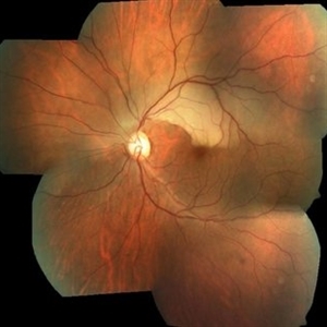
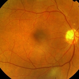
 Branch Retinal Artery Occlusion
Sep 21 2012 by Allen Chiang, MD, FASRS
Fundus photograph of a 27-year old male with a branch retinal artery occlusion. Systemic medical evaluation identified anti-phospholipid antibody syndrome.
Imaging device: Topcon
Condition/keywords: antiphospholipid antibody syndrome, branch retinal artery occlusion (BRAO)
-
 BRAO Rianto Color
Apr 12 2014 by Sjakon G Tahija, MD
Color image of the same patient.
Photographer: Avris Siahaan
Imaging device: Topcon
Condition/keywords: branch retinal artery occlusion (BRAO)
-
 Branch Retinal Artery Occlusion - FA
Sep 21 2012 by Allen Chiang, MD, FASRS
Fluorescein angiogram of a 27-year old male with a branch retinal artery occlusion demonstrates interruption of arterial flow and retrograde venous filling. Systemic medical evaluation identified anti-phospholipid antibody syndrome.
Imaging device: Topcon
Condition/keywords: antiphospholipid antibody syndrome, branch retinal artery occlusion (BRAO)
-
 AIDS - BAOI
Apr 8 2013 by Howard Schatz, MD
III AIDS - BAOI (BAO-AIDS)
Condition/keywords: AIDS, branch retinal artery occlusion (BRAO)
-
 Arterial Occlusion
Jul 14 2013 by Jason S. Calhoun
Patient in with vision loss in the lower quadrant of his visual field. Fundus photo shows arterial occlusion superior to the optic nerve
Photographer: Jason S. Calhoun, Department of Ophthalmology, Mayo Clinic Jacksonville, Florida
Imaging device: TOPCON TRC 50-EX
Condition/keywords: branch retinal artery occlusion (BRAO)
 Loading…