Search results (187 results)
-
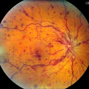
 Blistered Retina
Jan 27 2024 by prathibha hande, MS DNB
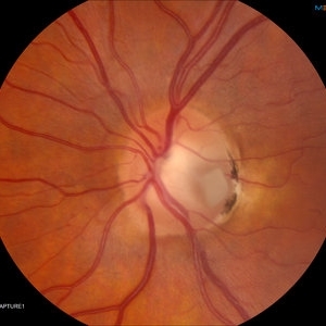
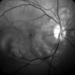
Fundus photo of a 32 year old male presenting with blurred vision. Undiagnosed renal hypertension. Blood pressure at the time of presentation 210/120 mmhg.
Photographer: Mr Prathap K
Imaging device: Mirante SLO fundus camera
Condition/keywords: hypertensive choroidopathy
-
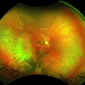
 Choroidal Detachment
Jan 17 2022 by Logan ryzenga
Left ultra-wide field photograph of an 81-year old female with a choroidal detachment affecting her left eye. Patient had a stent placed November, 2021 and following the procedure she complains of variable blurred vision and severe constricted visual fields. She presented at our office with flashes a month prior but without pain or floaters.
Photographer: Logan Ryzenga
Imaging device: Optos California
Condition/keywords: choroidal detachment, fundus photograph, left eye, Optos, pseudocolor, superior retina, ultra-wide field imaging
-
 Chorioretinitis with Overlying Vitreous Stranding/Vitritis
Mar 23 2023 by Isaac Agranoff
Fundus photograph of a 37-year-old woman presenting with chorioretinitis with overlying vitreous stranding/vitritis that has remained unchanged for multiple years. Patient presented with irritation and blurred vision and her vision was 20/40 OD. The OCT revealed evidence of low-grade inflammation and the recommend treatment was anti-inflammatory eye drops at this time and to obtain second opinion with another physician in the office.
Photographer: Isaac Agranoff, Technician
Imaging device: Optos California
Condition/keywords: chorioretinal scar, chorioretinitis, inflammation, Optos, ultra-wide field imaging, vitritis
-
 Central Retinal Vein Occlusion
Mar 27 2014 by Jason S. Calhoun
Patient in with mild blurred vision in the right eye. Fundus exam shows CRVO with scattered retinal hemorrhages in the right eye.
Photographer: Jason S. Calhoun, Mayo Clinic Jacksonville, Department of Ophthalmology
Imaging device: TOPCON TRC 50-EX
Condition/keywords: central retinal vein occlusion (CRVO)
-
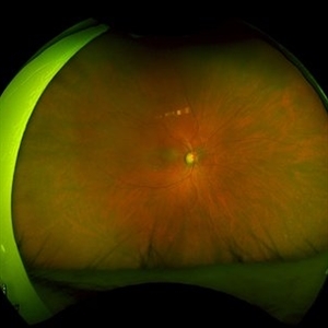
 Choroidal Metastasis
Apr 11 2024 by Corey Grant
Ultra-Widefield fundus photography and fundus autofluorescence images of a 61 year old female with Choroidal Metastasis affecting both eyes. Patient presented with blurred vision and flashes for a few weeks. Patient visual acuity was cc20/100 PH20/60 in the right eye and cc20/200 in the left eye. Patient admits to history of smoking for many years bit no known history of cancer prior to the visit. Physician recommended going to the ER for full body PET CT and stated that the first line of treatment is usually systemic chemo therapy. Patient will be reassessed in one month.
Photographer: Corey Grant
Imaging device: OPTOS CALIFORNIA RGB
Condition/keywords: cancer, choroidal metastasis, fundus autofluorescence (FAF), fundus photography, hyperautofluorescence, hypoautofluorescence, Optos, OPTOS CALIFORNIA RGB, Retina, ULTRA WIDE FIELD
-
 Choroidal Rupture
Sep 30 2023 by Jacob D. Grodsky, MD
24 year old female who presented after being hit in the head with a metal softball bat after an altercation. The patient reported blurred vision as well as a zig-zag line described as a “lightning strike” across her vision. Examination was significant for a choroidal rupture OD as well as commotio retinae OU.
Condition/keywords: choroidal rupture, commotio retinae, trauma
-
 Cilioretinal Artery Occlusion
May 14 2024 by Eloy Mata-Cortes, MD
Color image capturing the left eye of a 32-year-old female. Despite a negative ophthalmological and medical history, she reported three days of blurred vision and a paracentral scotoma in her left eye, while maintaining central vision. The image reveals retinal whitening, extends from the parafoveal region to the inferotemporal arcade indicative of cilioretinal artery occlusion. Following this observation, the patient was referred for systemic assessment to explore the underlying etiology of the occlusion.
Photographer: Eloy Mata-Cortes, MD, Instituto Mexicano de Oftalmología, Querétaro, México
Imaging device: Nidek Mirante
Condition/keywords: cilioretinal artery occlusion, oclussion, retinal whitening
-
 Congenital Retinal Macrovessel
Oct 13 2023 by Jacob D. Grodsky, MD
41 y/o male who presented with acute onset of blurred vision OD. Visual acuity was 20/200 OD; 20/25 OS. Examination was consistent with congenital retinal macrovessel through the macula with intraretinal hemorrhage as seen in the fundus photo. Intravitreal bevacizumab was injected, and visual acuity improved to 20/40 at 4-week follow-up. MRA head and neck was ordered to rule out other vascular anomalies.
Condition/keywords: congenital retinal macrovessel, RETINAL MACROVESSEL
-
 Dislocated Lens
Jun 29 2013 by Jason S. Calhoun
84-year-old female comes in with blurred vision in the left eye. VA was 20/30, right eye and count fingers in the left eye. Fundus examination reveals dislocation of the IOL into the vitreous inferiorily at 6-o'clock. Suggest surgery to fix the problem.
Photographer: Jason S. Calhoun, Mayo Clinic Jacksonville, Florida
Imaging device: TOPCON TRC 50-EX
Condition/keywords: dislocated posterior chamber intraocular lens (PCIOL)
-
 Hydroxychloroquine Maculopathy
Jul 23 2023 by Ahmad B. Tarabishy, MD
62 year old female with rheumatoid arthritis, treated with hydroxychloroquine 200 mg BID for the past 6-8 years. She presents with blurred vision, difficulty reading, and difficulty transitions from dark to light conditions since 4 months.
Photographer: Dr. Angela Rico
Condition/keywords: hydroxychloroquine toxicity, plaquenil toxicity, toxic maculopathy
-
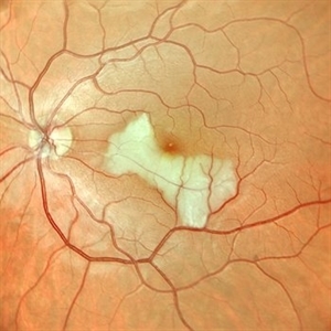
 Optic Nerve Pit With Sub-Retinal Fluid
Sep 17 2015 by Jason S. Calhoun
Young female with blurred vision in the left eye. Fundus photograph shows optic nerve pit adjacent to the macula where there is sub retinal fluid visible.
Photographer: Jason Calhoun, Mayo Clinic, Department of Ophthalmology
Imaging device: TOPCON-TRC50EX
Condition/keywords: congenital optic nerve pit
-
 Preeclampsia in a 30-Year-Old - Red Free Photograph - RE
Nov 25 2015 by Roy Schwartz, MD
A 30-year-old presented with central scotoma and blurred vision a day following C-section for preeclampsia.
Photographer: Galit Yair Pur
Condition/keywords: blurred vision, central scotoma, preeclampsia
-
 Racemose Angioma
Jan 23 2025 by SHILPI H NARNAWARE, ICO ( Retina) , FAICO ( Vitreo-Retina)
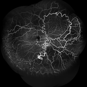
42 year old male, presented with blurred vision . Examination revealed Racemose angioma. FFA was done which revealed tortuosity of blood vessels.
Photographer: Shilpi Narnaware, Sarakshi Netralaya , Nagpur, Maharashtra , India
Imaging device: Mirante ( by Nidek)
Condition/keywords: FFA in a case of Racemose angioma
-
 Accordioning Crystalline Lens
Jul 8 2013 by Jason S. Calhoun
71-year-old male complained of blurred vision in the left eye. VA 20/40, right eye and 20/400, left eye without correction. Slit lamp exam shows Crystalline lens in both eyes. Right eye IOL is aligned and centered. Left eye shows an accordion of the crystalline lens. Retro illumination shows the IOL bent inward in the left eye. There was a 6-diopter difference of astigmatism between the right and left eye. Patient will have surgery to correct the issue.
Photographer: Jason S. Calhoun, Department of Ophthalmology, Mayo Clinic Jacksonville, Florida
Condition/keywords: dislocated crystalline lens
-
---thumb.JPG/image-square;max$300,300.ImageHandler) Accordioning Crystalline Lens
Accordioning Crystalline Lens
Jul 8 2013 by Jason S. Calhoun
71-year-old male complained of blurred vision in the left eye. VA 20/40, right eye and 20/400, left eye without correction. Slit lamp exam shows Crystalline lens, both eyes. Right eye IOL is aligned and centered. Left eye shows an accordion of the crystalline lens. Retro illumination shows the IOL bent inward in the left eye. There was a 6-diopter difference of astigmatism between the right and left eye. Patient will have surgery to correct the issue.
Photographer: Jason S. Calhoun, Department of Ophthalmology, Mayo Clinic Jacksonville, Florida
Condition/keywords: dislocated crystalline lens
-
---thumb.JPG/image-square;max$300,300.ImageHandler) Accordioning Crystalline Lens
Accordioning Crystalline Lens
Jul 8 2013 by Jason S. Calhoun
71-year-old male complained of blurred vision in the left eye. VA 20/40, right eye and 20/400, left eye without correction. Slit lamp exam shows Crystalline lens in both eyes. Right eye IOL is aligned and centered. Left eye shows an accordion of the crystalline lens. Retro Illumination shows the IOL bent inward in the left eye. There was a 6-diopter difference of astigmatism between the right and left eye. Patient will have surgery to correct the issue.
Photographer: Jason S. Calhoun, Department of Ophthalmology, Mayo Clinic Jacksonville, Florida
Condition/keywords: dislocated crystalline lens
-
---thumb.jpg/image-square;max$300,300.ImageHandler) Acute Blurred Central Vision
Acute Blurred Central Vision
Nov 7 2013 by Maurice F. Rabb
30 year old female with acute blurred central vision.
Condition/keywords: blurred vision
-
---thumb.jpg/image-square;max$300,300.ImageHandler) Acute Blurred Central Vision
Acute Blurred Central Vision
Nov 7 2013 by Maurice F. Rabb
30 year old female with acute blurred central vision.
Condition/keywords: blurred vision
-
---thumb.jpg/image-square;max$300,300.ImageHandler) Acute Blurred Central Vision
Acute Blurred Central Vision
Nov 7 2013 by Maurice F. Rabb
30 year old female with acute blurred central vision.
Condition/keywords: blurred vision
-
---thumb.jpg/image-square;max$300,300.ImageHandler) Acute Blurred Central Vision
Acute Blurred Central Vision
Nov 7 2013 by Maurice F. Rabb
30 year old female with acute blurred central vision.
Condition/keywords: blurred vision
-
---thumb.jpg/image-square;max$300,300.ImageHandler) Acute Posterior Placoid Pigment Epitheliopathy
Acute Posterior Placoid Pigment Epitheliopathy
Feb 4 2014 by Maurice F. Rabb
White female patient with 20/200 visual acuity in each eye with correction. Previously the patient had complained of a sore throat, headache associated with neck pain and mild nausea for several days. One week later, the patient stated she had blurred vision. There are no systemic diseases in the patient's past medical history. This is a possible case of acute posterior placoid pigment epitheliopathy.
Condition/keywords: acute posterior multifocal placoid pigment epitheliopathy (APMPPE)
-
 Aphakic Retinal Detachment
Nov 9 2012 by Norman Byer
This 75-year-old woman had a scleral buckling operation on this aphakic eye two months previously. Two months following surgery she again had new symptoms of blurred vision and was found to have a redetachment caused by this small tractional retinal tear which may have been missed at the time of her original operation.
Condition/keywords: aphakic eye, scleral buckle, tractional retinal tear
-
 Bilateral Macular Star
Mar 27 2014 by Jason S. Calhoun
Young female patient in with blurred vision in both eyes. VA is 20/40 in both eyes. Fundus photos show visible macular star centrally in both eyes. This is a result of Bilateral Neuroretinitis due to cat scratch.
Photographer: Jason S. Calhoun, Mayo Clinic Jacksonville, Department of Ophthalmology
Imaging device: TOPCON TRC 50-EX
Condition/keywords: macular star, neuroretinitis
-
 Bilateral Macular Star
Mar 27 2014 by Jason S. Calhoun
Young female patient in with blurred vision in both eyes. VA is 20/40 in both eyes. Fundus photos show visible macular star centrally in both eyes. This is a result of bilateral neuroretinitis due to cat scratch.
Photographer: Jason S. Calhoun, Mayo Clinic Jacksonville, Department of Ophthalmology
Imaging device: TOPCON TRC 50-EX
Condition/keywords: macular star, neuroretinitis
 Loading…