Search results (132 results)
-
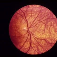
 Albinotic Fundus
Jan 24 2024 by Poornachandra B, MS, FVRS
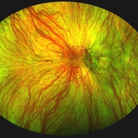
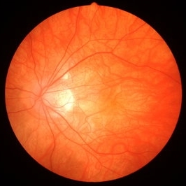
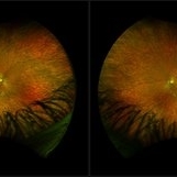
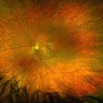
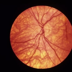
Fundus photo of a 30 year old male with Ocular albinism. Hypopigmented fundus with very evident choroidal vessels.
Photographer: Dr Poornachandra B
Condition/keywords: ocular albinism
-
 Albinotic fundus
Aug 10 2019 by Manish Nagpal, MD, FRCS (UK), FASRS
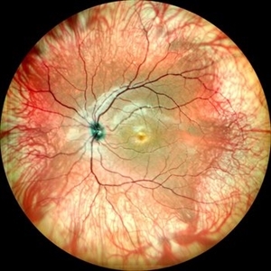
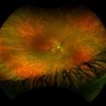
Albinotic fundus
Photographer: Gayathri Mohan, Retina Foundation
Imaging device: Nidek Mirante SLO
Condition/keywords: albinism
-
 Albinotic Fundus
Oct 1 2018 by Rameez N Hussain, MD
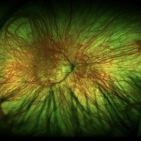
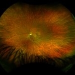
Albinotic fundus
Photographer: THAMBI DURAI (Edited by Lafas DE)
Imaging device: TOPCON
Condition/keywords: ocular albinism, oculocutaneous albinism
-
 Flat Fovea in Oculocutaneous Albinism
Oct 24 2020 by Guilherme Daher
Optical coherence tomography of a patient with oculocutaneous albinism showing a flat fovea.
Photographer: Jefferson Rocha, Instituto Suel Abujamra, Sao Paulo Brazil
Imaging device: Zeiss Cirrus HD-OCT 5000
Condition/keywords: albinism, fovea, foveal hypoplasia, nystagmus, oculocutaneous albinism, optical coherence tomography (OCT)
-
 Foveal Hypoplasia / Ocular Albinism
Aug 25 2024 by César Adrián Gómez Valdivia, MD
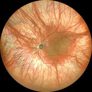
Fundus photograph of a 6-year-old female patient with foveal hypoplasia, ocular albinism and pendular nystagmus. Findings were bilateral. Retinal and Choroidal vasculature are exquisitely beautiful.
Photographer: @eyemissu2
Imaging device: TOPCON TRC-50DX
Condition/keywords: albinism, foveal hypoplasia, ocular albinism
-
 Fundus Changes of Albinism
Sep 20 2018 by Pengyao Lin
Fundus changes of albinism.
Condition/keywords: albinism, fundus photograph
-
 Fundus Changes of Albinism
Sep 20 2018 by Pengyao Lin
Fundus changes of albinism.
Condition/keywords: albinism, fundus photograph
-
 Grouped Albinotic Spots in Macula
Mar 4 2014 by David Callanan, MD
39-year-old female, 20/15 OU; normal color, ERG, EOG, thresholds, VF.
Condition/keywords: albinism, macula
-
 Iris in Albinism
May 1 2020 by Anfisa Ayalon, MD
Slit-lamp photograph of an 34-year-old man with oculocutaneous albinism. Note iris transillumination defects. Snellen chart visual acuity in both eyes-6/60. Nystagmus is also present.
Photographer: Anfisa Ayalon, MD., Meir Medical Center, Kfar Saba, Israel.
Condition/keywords: iris, nystagmus, oculocutaneous albinism, transillumination
-
 Iris Transillumination Defects in Albinism
May 1 2020 by Anfisa Ayalon, MD
Slit-lamp photograph of an 34-year-old man with oculocutaneous albinism. Note iris transillumination defects.
Photographer: Anfisa Ayalon, MD., Meir Medical Center, Kfar Saba, Israel.
Condition/keywords: albinism, iris, oculocutaneous albinism, transillumination
-
 Ocular Albinism
Aug 14 2021 by Aditya S Kelkar, MS, FRCS, FASRS,FRCOphth
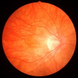
Fundus photograph of the left eye of a 26-year-old young man with ocular albinism.
Photographer: Devesh Kumar Dahariya, National Institute of Ophthalmology, Pune, India.
Imaging device: Zeiss Clarus 500
Condition/keywords: ocular albinism
-
 Ocular Albinism
Dec 11 2021 by Luis Daniel Gutierrez, MD
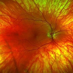
Optos images of 6-year-old female patient with type 1 oculocutaneous albinism.
Photographer: Luis Daniel Gutierrez García, Hospital Fundación Nuestra Señora de la Luz, Ciudad de México.
Imaging device: Optos
Condition/keywords: Albinism, ocular albinism, Optos, ultra-wide field imaging
-
 Oculo-cutaneous Albinisim
Aug 10 2025 by Akansha Sharma
Color fundus photograph of a 18 year old male with oculo-cutaneous albinisim.
Photographer: DR. AKANSHA SHARMA
Condition/keywords: albinism, foveal hypoplasia, oculocutaneous albinism
-
 Ocular Albinism
Mar 1 2014 by Homayoun Tabandeh, MD, FASRS
Ocular albinism and mild background diabetic retinopathy in a 27-year-old patient.
Condition/keywords: albinism, ocular albinism
-
 Oculocutaneous Albinism
Jan 22 2023 by Pietro Dechichi
Fundus photograph of a 6-year-old girl with oculocutaneous albinism. Patient's nystagmus made it difficult to perform the exam. Foveal hypoplasia and evident choroidal vessels can be seen in the retinography
Photographer: Pietro Dechichi
Imaging device: Optos California
Condition/keywords: childhood, foveal hypoplasia, ocular albinism
-
 Oculocutaneous albinism Slide 2
Oct 22 2012 by Ronald C. Gentile, MD
Fundus photo of the left eye revealed absence of retinal and choroidal pigmentation with foveal hypoplasia. The choroidal vasculature was more prominent then the retinal vasculature.
Photographer: The New York Eye & Ear Infirmary Department of Medical Imaging
Condition/keywords: choroidal pigmentation with foveal hypoplasia, choroidal vasculature, retinal vasculature
-
 "Mud-Splatter" of Posterior Pole and Peripheral Radial Streaks in a Carrier of Ocular Albinism
Jan 22 2019 by John S. King, MD
14-year-old healthy white female with family history of ocular albinism was seen by Dr. Hruby for a second opinion. Father and some of his brothers were positive for a history of ocular albinism. Va cc 20/30 J1+ OU; no nystagmus; no TIDs; no foveal hypoplasia. A "mud-spatter" appearance to the posterior pole was present, along with peripheral alternating streaks (photo). Dr. Hruby agreed that this was most likely a carrier of Ocular Albinism Type-1 (XR; GPR143 mutation), and possible genetic testing/counselling was discussed.
Photographer: Gretchen Harper
Imaging device: Optos California
Condition/keywords: Nettleship-Falls ocular albinism, ocular albinism
-
 "Mud-Splatter" of Posterior Pole and Peripheral Radial Streaks in a Carrier of Ocular Albinism
Jan 22 2019 by John S. King, MD
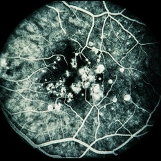
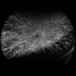
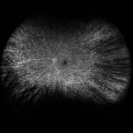
14-year-old healthy white female with family history of ocular albinism was seen by Dr. Hruby for a second opinion. Father and some of his brothers were positive for a history of ocular albinism. Va cc 20/30 J1+ OU; no nystagmus; no TIDs; no foveal hypoplasia. A "mud-spatter" appearance to the posterior pole was present, along with peripheral alternating streaks, which are very prominent in this late phase FA of the right eye. Dr. Hruby agreed that this was most likely a carrier of Ocular Albinism Type-1 (XR; GPR143 mutation), and possible genetic testing/counselling was discussed.
Photographer: Gretchen Harper
Imaging device: Optos California
Condition/keywords: Nettleship-Falls ocular albinism, ocular albinism
-
 "Mud-Splatter" of Posterior Pole and Peripheral Radial Streaks in a Carrier of Ocular Albinism
Jan 22 2019 by John S. King, MD
14-year-old healthy white female with family history of ocular albinism was seen by Dr. Hruby for a second opinion. Father and some of his brothers were positive for a history of ocular albinism. Va cc 20/30 J1+ OU; no nystagmus; no TIDs; no foveal hypoplasia. A "mud-spatter" appearance to the posterior pole was present, along with peripheral alternating streaks that are very prominent on this late phase FA OS. Dr. Hruby agreed that this was most likely a carrier of Ocular Albinism Type-1 (XR; GPR143 mutation), and possible genetic testing/counselling was discussed.
Photographer: Gretchen Harper
Imaging device: Optos California
Condition/keywords: Nettleship-Falls ocular albinism, ocular albinism
-
 "Mud-Splatter" of Posterior Pole and Peripheral Radial Streaks in a Carrier of Ocular Albinism
Jan 22 2019 by John S. King, MD
14-year-old healthy white female with family history of ocular albinism was seen by Dr. Hruby for a second opinion. Father and some of his brothers were positive for a history of ocular albinism. Va cc 20/30 J1+ OU; no nystagmus; no TIDs; no foveal hypoplasia. A "mud-spatter" appearance to the posterior pole was present, along with peripheral alternating streaks (photo). Dr. Hruby agreed that this was most likely a carrier of Ocular Albinism Type-1 (XR; GPR143 mutation), and possible genetic testing/counselling was discussed.
Photographer: Gretchen Harper
Imaging device: Optos California
Condition/keywords: Nettleship-Falls ocular albinism, ocular albinism
-
 "Mud-Splatter" of Posterior Pole and Peripheral Radial Streaks in a Carrier of Ocular Albinism
Jan 22 2019 by John S. King, MD
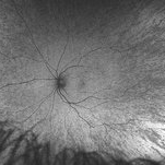
14-year-old healthy white female with family history of ocular albinism was seen by Dr. Hruby for a second opinion. Father and some of his brothers were positive for a history of ocular albinism. Va cc 20/30 J1+ OU; no nystagmus; no TIDs; no foveal hypoplasia. A "mud-spatter" appearance to the posterior pole was present, along with peripheral alternating streaks. Hypoautofluorescent areas correspond to hyperpigmented areas of retinal pigment epithelium, and vice versa (see photo). Dr. Hruby agreed that this was most likely a carrier of Ocular Albinism Type-1 (XR; GPR143 mutation), and possible genetic testing/counselling was discussed.
Photographer: Gretchen Harper
Imaging device: Optos California
Condition/keywords: Nettleship-Falls ocular albinism, ocular albinism
-
 Albinism
-
 Albinism
 Loading…