Search results (226 results)
-
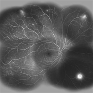
 Hemangioma Capilar Retina
Apr 9 2023 by Gustavo Aguirre-Suarez
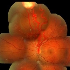
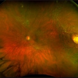
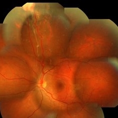
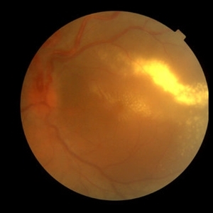
Fundus photograph composition of a Retinal Capilar Hemangioma
Photographer: Dr. Gustavo Aguirre-Suarez
Imaging device: Visucam 500
Condition/keywords: hemangioma, Von Hippel-Lindau
-
 VHL With Capillary Hemangioma Pre-Rx
Dec 29 2016 by Manish Nagpal, MD, FRCS (UK), FASRS
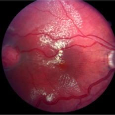
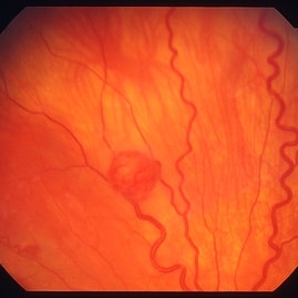
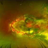
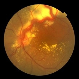
VHL with hemangioma with feeder vessels.
Photographer: rakesh juneja
Condition/keywords: cryotherapy, hemangioma, laser, Von Hippel-Lindau
-
 Von Hippel-Lindau Syndrome
Jul 12 2017 by Gabriel Costa Andrade, PhD
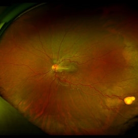
Fundus photograph of an 21-year-old girl with Von Hippel-Lindau Syndrome.
Photographer: Dr Gabriel Costa de Andrade
Imaging device: Optos® California
Condition/keywords: Von Hippel-Lindau
-
 Capillary Hemangioma
Dec 14 2016 by Young Hee Yoon, MD, PhD
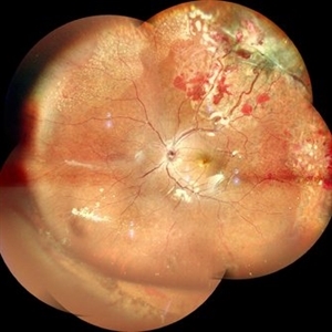
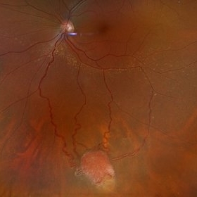
Wide fundus photo of a 35-year-old man with huge capillary hemagioma in the right eye. He is diagnosed with Von Hippel-Lindau disease. His best-corrected visual acuity was 20/50.
Photographer: Yu Jin Jang and Hun Eui Hong, Asan Medical Center
Imaging device: Wide fundus camera
Condition/keywords: retinal capillary hemangioma, Von Hippel-Lindau
-
 Solitary Retinal Capillary Hemangioblastoma
Apr 29 2019 by Michael A. Novak, MD
17-year-old young lady presented with reduced vision OD for several months. Her vision was 20/50.
Condition/keywords: retinal capillary hemangioblastoma, Von Hippel-Lindau
-
 Von Hippel-Lindau Syndrome
Mar 12 2016 by Sjakon G Tahija, MD
Fundus photograph of a patient with Von Hipple Lindau Disease and retinal angiomas.
Photographer: Avris Siahaan, Klinik Mata Nusantara
Condition/keywords: Von Hippel-Lindau
-
 Hemangioma
Feb 9 2021 by Kim Barrett
66-year-old female with a history of thyroid and uterine cancer in her 30's. She has a family history of cancers also. Current VA 20/40-2 PH OS. Patient and doctor chose observation at this time with possible surgical intervention in the future. She also has a small Hemangioma temporally in the right eye. Von Hippel-Lindau is also suspected and genetic testing was suggested.
Photographer: Kim Barrett C.O.A. Retina Specialists of Michigan, Grand Rapids, MI
Imaging device: Optos California
Condition/keywords: cancer, genetic testing, Optos, retinal hemangioblastoma, Von Hippel-Lindau
-
 Hemangioma of Retina
Sep 11 2018 by Carolyn Daley
50 degree OCT imaging of a 20-year-old with multiple bilateral hemangiomas. Patient was diagnosed with Von Hippel-Lindau Syndrome.
Photographer: Carolyn Daley, Retina Specialists of Michigan
Imaging device: Heidelberg Spectralis
Condition/keywords: 50 degrees, edema, hemangioma, optical coherence tomography (OCT), Von Hippel-Lindau
-
 Retinal Angiomas In VHL
Dec 24 2012 by Roy D. Brod, MD
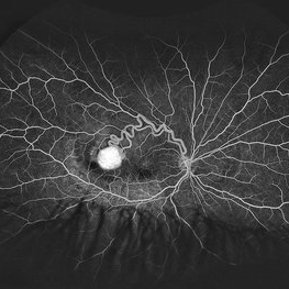
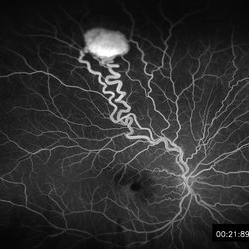
Mid phase fluorescein angiogram of 16 year old male with recent diagnosis of Von Hippel-Lindau disease showing hyperfluorescent angioma in superior mid periphery OD.
Photographer: Julia Walker
Condition/keywords: hemangioma, Von Hippel-Lindau
-
 Retinal capillary hemangiomas 3
Jan 11 2013 by Alex P. Hunyor, MD
Retinal capillary haemangiomas, left superior periphery, in a 20 year old female with von Hippel-Lindau disease.
Condition/keywords: hemangioma, retinal capillary hemangioma, Von Hippel-Lindau
-
 Retinal Hemangioblastoma Mid Phase FA
May 15 2013 by Robert T. Wendel, MD
20-year-old male. Genetic hx not yet defined.
Condition/keywords: Von Hippel-Lindau
-
 VHL Syndrome with Capillary Hemangioblastomas
Feb 26 2025 by Virginia Gebhart
39 year old female with choroidal hemangioma with capillary hemangioblastomas. Positive genetic testing for Von Hippel-Lindau Syndrome. Hemangioblastomas are stable compared to initial imaging in 2021. Pt started Welireg in Dec 2024, CNS tumors have started shrinking. No lesions in OD
Photographer: Virginia Gebhart, Retina Consultants of Carolina
Imaging device: Optos California
Condition/keywords: retinal capillary hemangioblastoma, Von Hippel-Lindau
-
 VHL With Capillary Hemangioma Post Cryo-Anti-VEGF
Dec 29 2016 by Manish Nagpal, MD, FRCS (UK), FASRS
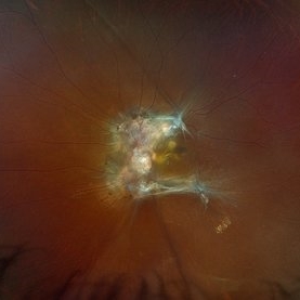
VHL with Haemangioma status post treatment with cryo and laser.
Photographer: Rakesh Juneja
Condition/keywords: cryotherapy, hemangioma, Von Hippel-Lindau
-
 Von Hippel Lindau Syndrome
Jun 9 2024 by Anjana Mirajkar, MS Ophthalmology
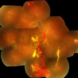
A widefield montage of a 23 year old female of LE case of VHL syndrome showing some hemorrhages with traction superiorly in a silicon oil filled eye with central settled retina. Cryo and laser marks are noted in periphery.
Photographer: Dr. Anjana Mirajkar -Retina Foundation, Ahmedabad
Imaging device: Mirante-Nidek
Condition/keywords: cryotherapy, exudative detachment, laser photocoagulation, vitreous hemorrhage, Von Hippel-Lindau
-
 Von Hippel-Lindau
Apr 3 2019 by Paola Brito, MD
30-year-old woman with Von Hippel-Lindau´s disease. Multiple hemangioblastoma. Big size tumors with macular involvement. Already received laser. She is being considered for surgical treatment
Photographer: Paola Brito, Hospital de la Luz, Mexico
Imaging device: Optos
Condition/keywords: Von Hippel-Lindau
-
 Von Hippel-Lindau
Sep 1 2014 by Hamid Ahmadieh, MD
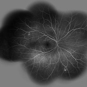
Two small retinal capillary hemangiomas detected by wide field FA in the symptom-free right eye of 30-year-old woman with Von Hippel-Lindau.
Photographer: Solmaz Shahmohammad, Negah Eye Center, Tehran, Iran
Condition/keywords: retinal capillary hemangioma, Von Hippel-Lindau
-
 Von Hippel-Lindau 1
Oct 13 2012 by Hamid Ahmadieh, MD
Color fundus photograph of the left eye of a 25-year-old woman with exudative retinal detachment secondary to retinal angiomatosis (Von Hippel-Lindau).
Photographer: Hamid Ahmadieh, MD, Ophthalmic Research Center, Labbafinejad Medical Center, Shahid Beheshti University of Medical Sciences
Imaging device: Topcon Fundus Camera
Condition/keywords: exudative retinal detachment, retinal angiomatous proliferation (RAP), Von Hippel-Lindau
-
 Von Hippel-Lindau Syndrome
Jul 23 2021 by Gabriel Costa Andrade, PhD
Fundus photograph of an 20-year-old woman with Von Hippel-Lindau Syndrome. The patient presented a ERM associated with vascular tumor in temporal periphery.
Photographer: Gabriel Andrade
Condition/keywords: tumor, Von Hippel-Lindau
-
 Von Hippel-Lindau Syndrome
Jan 7 2025 by Jordyn Beckman
Fundus photograph of an 37 year old female presents with reddish vascular lesion with feeder vessels for possible Von Hippel-Lindau Syndrome.
Photographer: Jordyn Beckman
Imaging device: California Optos
Condition/keywords: color fundus photograph, feeder vessel, genetic disorder, lesion, pre-cryotherapy
-
 Von Hippel Lindau
Mar 13 2013 by Carl C. Awh, MD, FASRS
TRD under silicone oil due to hemangiomas due to Von Hippel Lindau.
Photographer: Alecia Camp, CRA - Tennessee Retina - Nashville, TN
Condition/keywords: silicone oil, tractional retinal detachment, Von Hippel-Lindau
-
 Von Hippel-Lindau
Sep 3 2012 by Hamid Ahmadieh, MD
Color fundus photograph of a 35-year-old woman with retinal angiomatosis.
Photographer: Hamid Ahmadieh, MD, Ophthalmic Research Center, Labbafinejad Medical Center, Shahid Beheshti University of Medical Sciences
Imaging device: Topcon Fundus Camera
Condition/keywords: retinal angiomatous proliferation (RAP), Von Hippel-Lindau
-
 Retinal Angiomas In VHL
Dec 24 2012 by Roy D. Brod, MD
Fundus photograph of 16 year old male with recent diagnosis of Von Hippel-Lindau disease showing 2 retinal angiomas in inferior mid periphery OD.
Photographer: Julia Walker
Condition/keywords: hemangioma, Von Hippel-Lindau
-
 Von Hippel-Lindau
Oct 13 2012 by Hamid Ahmadieh, MD
Wide field FA image of the right eye of a 25-year-old woman with retinal angiomatosis (Von Hippel-Lindau). Fundus of the right eye seemed to be normal in ophthalmoscopy.
Photographer: Soodabeh Fooladin, Negah Eye Center, Tehran
Imaging device: Heidelberg Spectralis
Condition/keywords: exudative retinal detachment, retinal angiomatous proliferation (RAP), Von Hippel-Lindau
-
 Von Hippel-Lindau
Aug 23 2012 by Gabriela Lopezcarasa Hernandez, MD
29-year-old woman with decrease in visual acuity secondary to serous retinal detachment in Von Hippel-Lindau.
Photographer: Gabriela Lopezcarasa Hernandez, Hospital Angeles Lomas
Imaging device: FF4
Condition/keywords: serous retinal detachment
-
 Retinal Angiomas In VHL
Dec 24 2012 by Roy D. Brod, MD
Fundus photograph of 16 year old male with recent diagnosis of Von Hippel-Lindau disease showing typical appearance of a retinal angioma in superior mid periphery OD. Note unrelated choroidal nevus above superior arcade.
Photographer: Julia Walker
Condition/keywords: hemangioma, Von Hippel-Lindau
 Loading…