Search results (59 results)
-
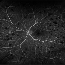
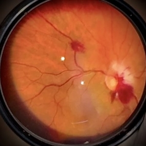
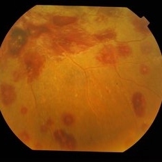
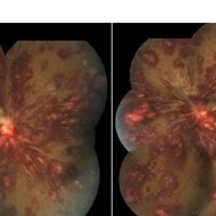
 Anaemic Retinopathy
Sep 13 2023 by Anand Temkar
Wide field image of the RE of a 35 year old male patient showing Roth's spots in all four quadrants and venous tortuosity in a case of Anaemic Retinopathy.
Photographer: Dr.Anand Temkar- Retina Foundation, Ahmedabad
Imaging device: Mirante
Condition/keywords: anaemic retinopathy, roth spots
-
 Leukemic Retinopathy
Oct 9 2012 by Sharon Fekrat, MD FACS FASRS
22-year-old female with new diagnosis of acute myelogenous leukemia. White blood cell count was 35,000,000,000 cells/L. Note Roth Spots.
Photographer: Tiffanie Keaton, Duke Eye Imaging, Durham, NC
Condition/keywords: acute leukemia, white centered retinal hemorrhage (Roth Spot)
-
 Roth Spots
Oct 26 2022 by Denica Rodriguez
Roth spots during optos FA on a 68 year old female with retinal hemorrhage effecting her left eye. Patient was referred for non-proliferative diabetic retinopathy without macular edema.
Photographer: Denica Rodriguez & Zachary Seim
Imaging device: Optos California
Condition/keywords: Diabetes, FLUORESCEIN ANGIOGRAPHY, left eye, Optos, Retina, Roth Spots, ultra-wide field imaging
-
 Arcus Retinalis
Jun 21 2025 by Moazzam Parvez
Fundus photograph of a 30 year oiled gentleman with multiple dome shaped sub hyaloid haemorrhage with discrete arches retinals around it. Roth spots are also noted on the retina.
Photographer: Moazzam Parvez , Netralayam , Kolkata
Imaging device: Topcon Maestro 2
Condition/keywords: arcus retinalis, Roth spots, Sub hyaloid haemorrhage
-
 Hypertensive Retinopathy
Dec 24 2017 by Purva Patwari
52-year-old female diagnosed of hypertension by retina evaluation.
Photographer: Dr Purva Patwari, Patwari Retina Center, Ahmedabad, Gujarat , India
Imaging device: ZEISS VISU500
Condition/keywords: hypertensive retinopathy, neovascularization elsewhere (NVE), Roth spots
-
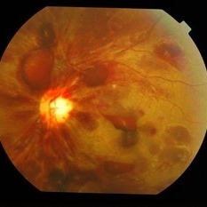
 Multiple Blot Hemorrhages and Roth Spots
Jan 24 2018 by Gabriel Costa Andrade, PhD
Multiple blot hemorrhages and Roth spots in a patient with acute leukemia.
Photographer: Gabriel Andrade, MD
Condition/keywords: leukemia, Roth spots
-
 Acute Myeloid Leukemia
Dec 4 2018 by Linda A Cernichiaro- Espinosa, MD
Fundus photograph of a 12-year-old girl with superficial and deep retinal hemorrhages associated to acute myeloid leukemia (AML). A subhyaloid bleed involves the macula in both eyes.
Photographer: Dr. Linda A Cernichiaro Espinosa
Imaging device: inView (Volk Inc. USA) with iPhone 6
Condition/keywords: acute leukemia, leukemia, retinopathy, Roth spots
-
 Dengue Fever
Oct 25 2012 by Mallika Goyal, MD
Fundus photograph of the left eye of a 32-year-old gentleman with dengue fever and thrombocytopenia. Photograph shows extensive retinal and pre-retinal haemorrhages, roth spots but no dengue retinitis. Same patient as in images 1-5.
Condition/keywords: Dengue Fever, preretinal hemorrhage, rosacea conjunctivitis
-
 Thrombocytopenia
Sep 24 2024 by DR Rohit Gupta
Fundus photography of a 16 year-old girl suffering from severe thrombocytopenia, showing flame shaped hemorrhage.
Photographer: Dr Rohit gupta
Imaging device: Samsung S21
Condition/keywords: anaemic retinopathy, flame shaped retinal hemorrhage, Haemorrhage, Roth spots, white centered retinal hemorrhage (Roth Spot), white dot syndrome
-
 Bilateral Roth Spot in the Setting of Mitral Valve Endocarditis
Aug 18 2025 by Helder Vasconcelos
A 55-year-old man with chronic alcoholism presented with wasting and fever. The symptoms were preceded by a recent tooth extraction and gingivitis. Fundus examination in the ICU showed a retinal hemorrhage with a white spot (Roth spot) associated with peripapillary hemorrhage and cotton wool exudate. A similar Roth spot was observed in the contralateral eye.
Photographer: Helder Vasconcelos
Imaging device: Smartphone Fundoscopy
Condition/keywords: Infectious endocarditis, Roth Spots
-
 Chronic Myelogenous Leukemia
May 27 2024 by Akansha Sharma
Color fundus photograph of a 41 year old male presenting with ocular manifestations of chronic myelogenous leukemia.
Photographer: Dr. Akansha Sharma, Bharati Eye Hospital
Condition/keywords: CML, Roth Spots
-
 Chronic Myelogenous Leukemia
May 27 2024 by Akansha Sharma
Color fundus photograph of a 41 year old male presenting with ocular manifestations of chronic myelogenous leukemia.
Photographer: Dr. Akansha Sharma, Bharati Eye Hospital
Condition/keywords: CML, Roth Spots
-
 Dengue Fever
Oct 25 2012 by Mallika Goyal, MD
Fundus photograph of the left eye of a 32-year-old gentleman with dengue fever and thrombocytopenia. Photograph shows extensive retinal and pre-retinal haemorrhages, roth spots but no dengue retinitis. Same patient as in images 1-5
Condition/keywords: Dengue Fever, preretinal hemorrhage, rosacea conjunctivitis
-
 Dengue Fever
Oct 25 2012 by Mallika Goyal, MD
Fundus photograph of the left eye of a 32-year-old gentleman with dengue fever and thrombocytopenia. Photograph shows extensive retinal and pre-retinal haemorrhages, roth spots but no dengue retinitis.
Condition/keywords: Dengue Fever, preretinal hemorrhage, rosacea conjunctivitis
-
 Dengue Fever
Oct 25 2012 by Mallika Goyal, MD
Fundus photograph of the right eye of a 32-year-old gentleman with dengue fever and thrombocytopenia. Photograph shows extensive retinal and pre-retinal haemorrhages, roth spots but no dengue retinitis. Same patient as in images 1-5.
Condition/keywords: Dengue Fever, rosacea conjunctivitis, thrombocytopenia
-
 Dengue Fever
Oct 25 2012 by Mallika Goyal, MD
Fundus photograph of the right eye of a 32-year-old gentleman with dengue fever and thrombocytopenia. Photograph shows extensive retinal and pre-retinal haemorrhages, roth spots but no dengue retinitis. Same patient as in images 1-5.
Condition/keywords: Dengue Fever, preretinal hemorrhage, rosacea conjunctivitis
-
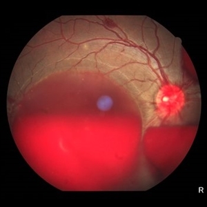
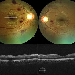 Dengue-Associated-Retinopathy (Anaemic Retinopathy)
Jan 16 2018 by Deepak Bhojwani, MS
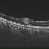
22-year-old male with systemic dengue fever and anaemia presenting with roth spots in both eyes (OD>OS). Horizontal raster OCT scans showing intraretinal foveal hameorrhage in right eye.
Photographer: Dr Deepak Bhojwani, Raghudeep Eye Hospital , Ahmedabad
Imaging device: Zeiss- HD- OCT
Condition/keywords: anaemic retinopathy, Roth spots
-
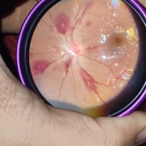
 Leukemic Infiltrate
May 11 2025 by Hemanth Murthy, MBBS, MD, FASRS
43 year male patient presented with blurring of vision in right eye since 3 days. Vision 6/12 and left eye vision was 6/6. Haematological workup showed Hemoglobin -10g/dl, WBC count 276440 cells/cu.mm Smear showed large immature myeloid cells.
Photographer: Mr Veda Vyas
Condition/keywords: Acute myeloid leukaemia with Roth spots and leukaemia infiltrates
-
 Leukemic Retinopathy
Apr 20 2019 by Jitendra Kumar
Fundus photograph of 27-year-old acute leukemic patient came to OPD with history of hand movement. Fundus photo shows diffuse haemorrheges with Roth spots in both eyes.
Photographer: DR JITENDRA KUMAR, SRI SANKARADEVA NETHRALAYA, GUWAHATI
Imaging device: Zeiss fundus camera
Condition/keywords: leukemia, Roth spots
-
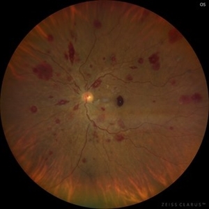 Leukemic Retinopathy
Nov 27 2024 by Ramses Rosales-Diaz
Fundus photograph of a 48-year-old woman with venous dilatation and tortuosity, flame-shaped and intraretinal hemorrhages, Roth spots and sub-ILM hemorrhage. Her complete blood count reports 425,540 lymphocytes/microliter, and the blood smear reveals Gumprecht shadows and numerous lymphocytes with nuclei exhibiting hypercondensed chromatin. She is diagnosed with chronic lymphocytic leukemia and receives appropriate treatment from the hematology team
Photographer: Ramses Rosales-Diaz, Asociación Para Evitar la Ceguera en México
Imaging device: Clarus 700
Condition/keywords: leukemia, sub ILM hemorrhage, white centered retinal hemorrhage (Roth Spot)
-
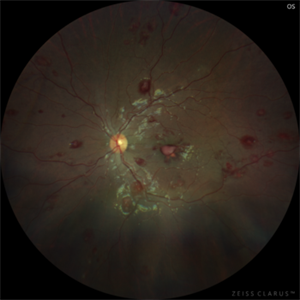
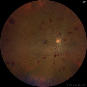 Leukemic Retinopathy
Nov 27 2024 by Ramses Rosales-Diaz
Fundus photograph of a 48-year-old woman showing venous dilatation and tortuosity, flame-shaped hemorrhages, intraretinal hemorrhages, sub-ILM hemorrhages, and Roth spots. Her complete blood count shows 425,540 lymphocytes/microliter, and the blood smear reveals Gumprecht shadows along with numerous lymphocytes with hypercondensed chromatin in their nuclei. She is diagnosed with chronic lymphocytic leukemia and receives appropriate treatment from the hematology team.
Photographer: Ramses Rosales-Diaz, Asociación Para Evitar la Ceguera en México
Imaging device: Zeiss Clarus 700
Condition/keywords: leukemia, sub ILM hemorrhage, white centered retinal hemorrhage (Roth Spot)
-
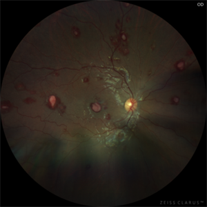
 Leukemic Retinopathy
Nov 27 2024 by Ramses Rosales-Diaz
Fundus photograph of a 48-year-old woman showing venous dilatation and tortuosity, flame-shaped hemorrhages, intraretinal hemorrhages, sub-ILM hemorrhages, and Roth spots. Her complete blood count shows 425,540 lymphocytes/microliter, and the blood smear reveals Gumprecht shadows along with numerous lymphocytes with hypercondensed chromatin in their nuclei. She is diagnosed with chronic lymphocytic leukemia and receives appropriate treatment from the hematology team.
Photographer: Ramses Rosales-Diaz, Asociación Para Evitar la Ceguera en México
Imaging device: Zeiss Clarus 700
Condition/keywords: leukemia, sub ILM hemorrhage, white centered retinal hemorrhage (Roth Spot)
-
 Leukemic Retinopathy - OD
Aug 1 2023 by Shaleen Arora
A 14-year-old female was transferred from an outside hospital with a new diagnosis of B-ALL and WBC of 667,000. Following lumbar puncture, she developed blurry vision and floaters but denied curtaining, flashes, and diplopia. Ophthalmology was consulted to assess for disc edema. Exam revealed visual acuity of 20/100 OD and 20/200 OS. Imaging showed diffuse hemorrhages and Roth spots OU, consistent with leukemic retinopathy. The patient was followed by retinal specialists with spontaneous improvement in visual acuity over three weeks.
Photographer: Camilo Martinez, Childrens National Medical Center, Department of Ophthalmology
Condition/keywords: leukemia, leukemic infiltration, retinopathy, Roth spots
-
 Leukemic Retinopathy - OS
Aug 1 2023 by Shaleen Arora
A 14-year-old female was transferred from an outside hospital with a new diagnosis of B-ALL and WBC of 667,000. Following lumbar puncture, she developed blurry vision and floaters but denied curtaining, flashes, and diplopia. Ophthalmology was consulted to assess for disc edema. Exam revealed visual acuity of 20/100 OD and 20/200 OS. Imaging showed diffuse hemorrhages and Roth spots OU, consistent with leukemic retinopathy. The patient was followed by retinal specialists with spontaneous improvement in visual acuity over three weeks.
Photographer: Camilo Martinez, Childrens National Medical Center, Department of Ophthalmology
Condition/keywords: leukemia, leukemic infiltration, retinopathy, Roth spots
-
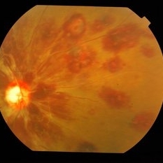
 Macular Hemorrhage Secondary to Anemic Retinopathy
Apr 18 2022 by Deepak Bhojwani, MS
Fundus image of a young 28 year old patient who has been diagnosed as 'PRIMARY BONE MARROW APLASIA' by hematologist showing large macular hemorrhage (sub -ILM Heme mound). Few Roth spots were also seen in midperiphery suggesting 'ANEMIC RETINOPATHY'.
Photographer: DEEPAK BHOJWANI
Condition/keywords: anaemic retinopathy, BONE MARROW APLASIA
 Loading…