Search results (38 results)
-
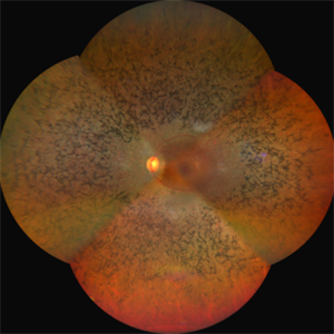
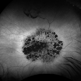
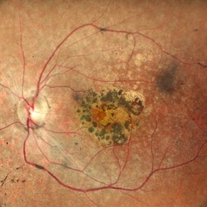
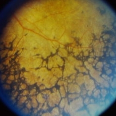
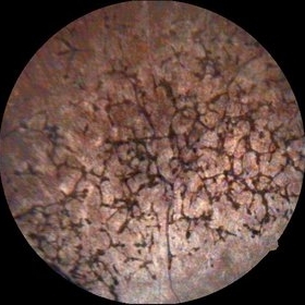 Bone Corpuscle Pigments
Sep 11 2014 by Mehul A Shah
A 42-year-old female presented with gradual reduction in vision.
Photographer: Drashti Netralaya,Dahod
Imaging device: FF 450
Condition/keywords: retinitis pigmentosa (RP) dystrophy
-
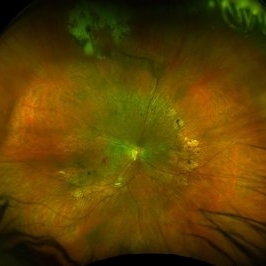
 Mucopolysaccharidosis Type III
Apr 21 2023 by Matthew Dombrow, MD
Fundus photograph and autofluorescence of a 49 year old male with mucopolysaccharidosis type III (Sanfilippo syndrome)
Photographer: Cori Sturtevant, Connecticut Retina Consultants, Hamden, Connecticut
Condition/keywords: mucopolysaccharidoses, retinitis pigmentosa (RP) dystrophy
-
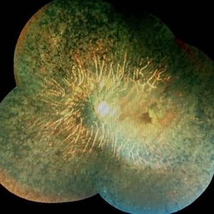
 Mucopolysaccharidosis Type III (Sanfilippo syndrome)
Apr 21 2023 by Matthew Dombrow, MD
Fundus photograph and autofluorescence of a 49 year old male with mucopolysaccharidosis type III (Sanfilippo syndrome)
Photographer: Cori Sturtevant, Connecticut Retina Consultants, Hamden, Connecticut
Condition/keywords: mucopolysaccharidoses, retinitis pigmentosa (RP) dystrophy
-
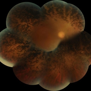
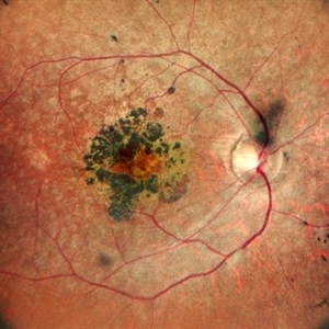
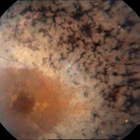 Retinitis Pigmentosa With Macular Involvment
Sep 11 2014 by Mehul A Shah
A 35-year-old female presented with gradual reduction in vision.
Photographer: Drashti Netralaya,Dahod
Imaging device: FF 450
Condition/keywords: retinitis pigmentosa (RP) dystrophy
-
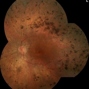
 Unilateral Retinitis Pigmentosa
May 1 2014 by Raj K. Maturi, MD
53-year-old woman with significant salt and pepper retinopathy OS.
Photographer: Tom Steele, Midwest Eye Institute
Condition/keywords: retinitis pigmentosa (RP) dystrophy
-
 A Feast for Crows , Retinitis pigmentosa
Sep 22 2022 by wang xiaomei
Fundus photograph of an 55-year-old man with Retinitis Pigmentosa, There is increasing loss of pigment from the pigment epithelium with intraretinal clumping of melanin, appearing most often as coarse clumps in a "bone spicule" configuration, arteriolar narrowing
Photographer: Man, Li, Bao Ji Ophthalmic Hospital
Imaging device: ZEISS CLARUS 500
Condition/keywords: retinitis pigmentosa (RP) dystrophy
-
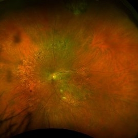
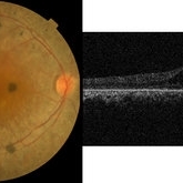
 Advanced Retinitis Pigmentosa
Mar 14 2017 by Hamid Ahmadieh, MD
Merged color fundus photograph of the right eye of a patient with advanced retinitis pigmentosa sparing the posterior pole.
Photographer: Soodabeh Fouladin, Negah Eye Center, Tehran, Iran
Condition/keywords: color fundus photograph, retinitis pigmentosa (RP) dystrophy
-
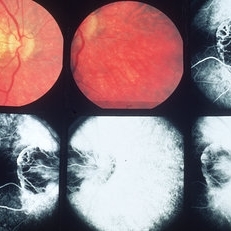 Inflammation
May 15 2013 by Howard Schatz, MD
30-year-old white female, 20/30 periphery and posterior RP dystrophy.
Condition/keywords: inflammation, retinitis pigmentosa (RP) dystrophy
-
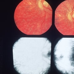 Inflammation
May 15 2013 by Howard Schatz, MD
30-year-old white female, 20/50 periphery and posterior RP dystrophy.
Condition/keywords: inflammation, retinitis pigmentosa (RP) dystrophy
-
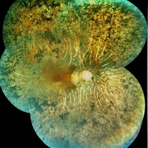
 Mucopolysaccharidosis Type III (Sanfilippo syndrome)
Apr 21 2023 by Matthew Dombrow, MD
Fundus photograph and autofluorescence of a 49 year old male with mucopolysaccharidosis type III (Sanfilippo syndrome)
Photographer: Cori Sturtevant, Connecticut Retina Consultants, Hamden, Connecticut
Condition/keywords: mucopolysaccharidoses, retinitis pigmentosa (RP) dystrophy
-
 Retinitis Pigmentosa
Apr 30 2015 by Mitzy E Torres Soriano, MD
Fundus of patient with retinitis pigments, bone spicule-shaped pigment deposits are present with retinal atrophy, while the macula is preserved . Retinal vessels are attenuated.
Photographer: Mitzy E. Torres Soriano, MD; Centro medico Cagua-Estado Aragua. Venezuela
Imaging device: TRC-NW8
Condition/keywords: pigmentary retinal dystrophy, retinal dystrophy, retinitis pigmentosa, retinitis pigmentosa (RP) dystrophy
-
 Retinitis Pigmentosa
Aug 25 2015 by René Hernán Parada Vásquez
Fundus photograph of both eyes of a 38-year-old female with retinitis pigmentosa, bone spicule-shaped pigment deposits are present in the mid periphery, and macula with a peripheral ring of depigmentation.
Photographer: Parada René, ESO, Guatemala.
Imaging device: Canon CR-2
Condition/keywords: bilateral pigmentary retinopathy, retinitis pigmentosa, retinitis pigmentosa (RP) dystrophy
-
 Retinitis pigmentosa
Feb 26 2020 by Manuel Ángel Alcántara Delgado, MD
Merged color fundus photograph of a 68-year-old woman with advanced retinitis pigmentosa. It is appreciated bone spicule-shaped pigment deposits, optic disc pallor, retinal atrophy and attenuated retinal vessels.
Photographer: Manuel Ángel Alcántara Delgado
Condition/keywords: choroidal circulation, optic disc pallor, pericentral retinitis pigmentosa, retina, retinitis pigmentosa, retinitis pigmentosa (RP) dystrophy, sector retinitis pigmentosa
-
 Retinitis Pigmentosa
Feb 26 2020 by Manuel Ángel Alcántara Delgado, MD
Merged color fundus photograph of a 68-year-old woman with advanced retinitis pigmentosa. It is appreciated bone spicule-shaped pigment deposits, optic disc pallor, retinal atrophy, attenuated retinal vessels and surface wrinkling retinopathy.
Photographer: Manuel Ángel Alcántara Delgado
Condition/keywords: chorioretinal atrophy, choroidal circulation, optic disc pallor, pericentral retinitis pigmentosa, retina, retinitis pigmentosa, retinitis pigmentosa (RP) dystrophy, sector retinitis pigmentosa
-
 RETINITIS PIGMENTOSA
Oct 15 2022 by Akansha Sharma
COLOUR FUNDUS PHOTOGRAPH OF A 30 YEAR OLD MALE WITH RETINITIS PIGMENTOSA
Photographer: Dr. Akansha Sharma-Retina Foundation, Ahmedabad
Condition/keywords: retinitis pigmentosa (RP) dystrophy, RP variant
-
 RETINITIS PIGMENTOSA
Oct 15 2022 by Akansha Sharma
COLOUR FUNDUS PHOTOGRAPH OF A 30 YEAR OLD MALE WITH RETINITIS PIGMENTOSA
Photographer: Dr. Akansha Sharma-Retina Foundation, Ahmedabad
Condition/keywords: retinitis pigmentosa (RP) dystrophy, RP variant
-
 Retinitis Pigmentosa
Oct 16 2024 by Virginia Gebhart
74 year old female with bone spicule pigmentation associated with Retinitis Pigmentosa. Pt diagnosed at age 53, relatively asymptomatic prior to diagnosis. Pt reports gradual vision loss over 10+ years. BCVA 20/40
Photographer: Virginia Gebhart, Retina Consultants of Carolina
Imaging device: Optos California
Condition/keywords: bone spicule, retinitis pigmentosa, retinitis pigmentosa (RP) dystrophy
-
 Retinitis Pigmentosa
Jan 15 2025 by Virginia Gebhart
52 year old male with advanced RP OU. BCVA HM OD, LP OS. Referred to genetic specialist per pt request to discuss gene therapy.
Photographer: Virginia Gebhart, Retina Consultants of Carolina
Imaging device: Optos California
Condition/keywords: bone spicule, retinitis pigmentosa, retinitis pigmentosa (RP) dystrophy
-
 Retinitis Pigmentosa
Jan 25 2024 by Virginia Gebhart
58 year old female diagnosed at age 13. Vision 20/100
Photographer: Virginia Gebhart
Imaging device: Topcon
Condition/keywords: retinitis pigmentosa, retinitis pigmentosa (RP) dystrophy
-
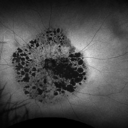
 Retinitis Pigmentosa
Oct 12 2023 by Virginia Gebhart
Fundus Auto-Fluorescence photo of 73-year-old woman with Retinitis Pigmentosa, first diagnosed 23 years ago. Extensive outer retinal atrophy with minimal foveal sparing, bone spicule pigmentation and waxy pallor. Vision NLP
Photographer: Virginia Gebhart, Retina Consultants of Carolina
Imaging device: Optos
Condition/keywords: retinitis pigmentosa, retinitis pigmentosa (RP) dystrophy
-
 Retinitis Pigmentosa & CME
Oct 10 2015 by Hamid Ahmadieh, MD
Color fundus photograph of the right eye of a 40-year-old woman with cystoid macular edema ( CME) secondary to retinitis pigmentosa (RP).
Photographer: Shabnam Pooreh, Negah Eye Center, Tehran, Iran
Condition/keywords: color fundus photograph, cystoid macular edema (CME), retinitis pigmentosa
-
 Retinitis Pigmentosa (RP)
Sep 21 2023 by Ben Serar
Fundus photograph showing bony spicules and arteriolar attenuation in a case of Retinitis Pigmentosa.
Condition/keywords: Retinitis Pigmentosa (RP)
-
 Retinitis Pigmentosa (RP)
Sep 21 2023 by Ben Serar
Fundus photograph showing bony spicules in a case of Retinitis Pigmentosa.
Condition/keywords: Retinitis Pigmentosa (RP)
-
 Retinitis Pigmentosa (RP)
Sep 14 2023 by Ben Serar
Fundus photograph showing bony spicules in a case of Retinitis Pigmentosa.
Condition/keywords: Bony spicules, Retinitis Pigmentosa (RP)
 Loading…