Search results (41 results)
-
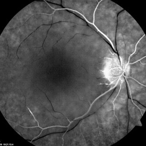
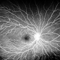
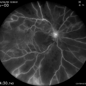
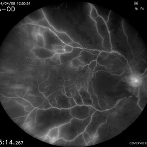
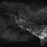
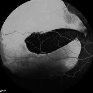
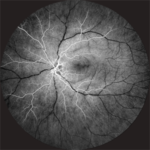 Takayasu Retinopathy
Apr 30 2025 by Vishal Agrawal, MD, FRCS,FACS,FASRS
Fundus fluorescein angiography image of a young girl with diagnosed Takayasu arteritis who presented with complains of diminished vision in both eyes. FFA shows complete absence of venous filling with segmented blood column secondary to CRAO with peripheral avascular area.
Photographer: Dr Ayushi Gupta
Imaging device: Clarus 700
Condition/keywords: calcified drusen, CRAO, takayasu arteritis
-
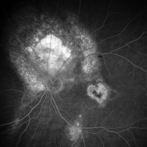
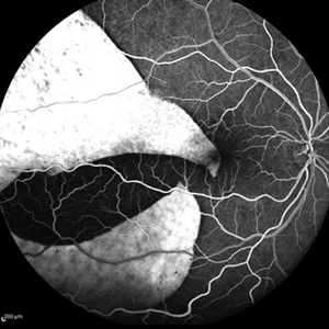 Giant RPE-rip
Sep 5 2021 by Hemanth Murthy, MBBS, MD, FASRS
Fundus fluorescein angiography of a 50 year-old patient with spontaneous giant RPE rip.
Photographer: Mr Veda Vyas
Imaging device: Heidelberg HRA
Condition/keywords: RPE-Rip
-
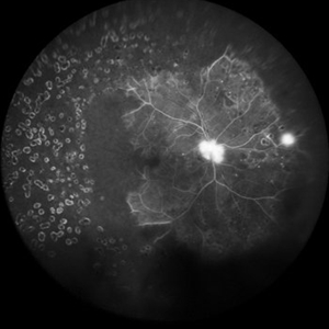
 Idiopathic retinal vasculitis, aneurysms and neuroretinitis
Apr 24 2022 by Aniruddha K Agarwal, MD
Ultra-wide field fundus fluorescein angiography (FFA) of the left eye from an asymptomatic, healthy 33-year-old woman who was referred to the retina clinic from a refractive surgery unit due to the presence of vascular anomalies and hard exudates in both eyes. FFA revealed the characteristic sacular aneurysms at the bifurcation of retinal arterioles in the posterior pole, together with microvascular anomalies and capillary closure peripherally.
Photographer: Julio J GONZALEZ-LOPEZ, MD, PhD, FEBO and Teresa GONZALEZ-LOMAS, RN
Imaging device: Optos California
Condition/keywords: IRVAN Syndrome, IUSG, neuroretinitis, retinal vasculitis, uveitis
-
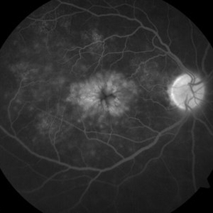
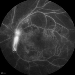 Optic Nerve Head Cannonball
Dec 15 2019 by Veer Singh, MS, FVRS, FMRF, FICO (Retina)
This is the fundus fluorescein angiography (FFA) of the left eye of a 62-year-old diabetic patient with proliferative diabetic retinopathy and neovascularization of disc who bled from the disc while he was undergoing an FFA procedure. The bleed from the disc gives the appearance of a cannonball fired from a cannon hence the caption "Optic Nerve Head Cannonball".
Photographer: Dr. Veer Singh
Imaging device: Heidelberg Spectralis HRA
Condition/keywords: fluorescein angiogram (FA), neovascularization of the disc (NVD), optic nerve head, proliferative diabetic retinopathy (PDR), vitreous hemorrhage
-
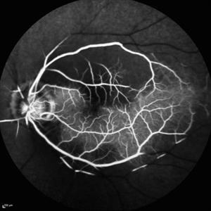
 Takayasu Retinopathy
Apr 30 2025 by Vishal Agrawal, MD, FRCS,FACS,FASRS
Fundus fluorescein angiography image of a young girl with diagnosed Takayasu arteritis who presented with complains of diminished vision in both eyes. FFA shows complete absence of venous filling with segmented blood column secondary to CRAO with peripheral avascular area.
Photographer: Dr Ayushi Gupta
Imaging device: Clarus 700
Condition/keywords: CRAO, Takayasus disease
-
 Central Retinal Artery Occlusion with Cilioretinal Sparing - Fundus Fluorescein Angiography
Oct 28 2020 by Fang Helen Mi
Fundus fluorescein angiogram confirmed a delayed arm-retinal time and extensive capillary fallout, with cilioretinal artery sparing.
Condition/keywords: central retinal artery occlusion (CRAO), cilioretinal sparing
-
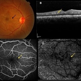 Paracentral Acute Middle Maculopathy
Oct 25 2019 by Gayathri Mohan
Multimodal images of a case of a 29-year-old female with paracentral acute middle maculopathy. A-color fundus photograph showing multiple confluent white retinal patches. B- On OCT the acute lesions of PAMM characteristically appear as placoid, hyperreflective bands at the level of the INL C-Fundus fluorescein angiography showing a capillary nonperfusion area D-flow void areas in deep capillary plexus
Photographer: Akshar Soni
Imaging device: Heidelberg, Nidek
Condition/keywords: fundus albipunctatus, optical coherence tomography (OCT), paracentral acute middle maculopathy
-
 Proliferative DR OD Final
Apr 28 2017 by SHRUTI CHANDRA, MD
Digital widefield fundus fluorescein angiography of a 56-year-old diabetic patient with visual acuity 6/18 and 6/6 in right and left eye respectively. This image depicts extensive peripheral capillary non perfusion with preserved posterior pole perfusion.
Photographer: Shruti Chandra
Imaging device: Heidelberg Spectralis
Condition/keywords: proliferative diabetic retinopathy (PDR)
-
 Branch Retinal Artery Occlusion
May 4 2021 by Priya Rasipuram Chandrasekaran, MBBS, DO, DNB, FRCS
This is the fundus photo of a 52-year-old male taken within 6 hours and after 24 hours of sudden onset of inferior field loss. The photo shows prominence of retinal edema in the region of arterial occlusion as time passes by. The optical coherence tomogram scan taken vertically through the normal and the involved area shows thickening and hyper reflectivity of retinal nerve fiber layer and decreased reflectivity of the retinal layers beneath it (white arrow). Fundus fluorescein angiography shows complete non-filling of the artery in the early phase with slow filling in the late phase and highlighting the embolus.
Condition/keywords: branch retinal artery occlusion (BRAO)
-
 BRVO-MCR-FFA
Jun 27 2025 by Gayathri M S
Case of impending Macular BRVO. 52 year old female on medication for Hypertension and Diabetes Mellitus since 2 years. BCVA 6/18,N6. IOP 16 mmHg. Multicolor Reflectance and Fundus Fluorescein Angiography picture shows mild dilated tortuous inferior vessels, small areas of capillary non perfusion and few microanurysms.
Photographer: Gayathri MS
Imaging device: Heidelberg spectralis
Condition/keywords: fluorescein angiogram (FA), macular branch retinal vein occlusion (BRVO), multicolor
-
 Central Retinal Artery Occlusion
Mar 2 2021 by Renata Garcia Franco, Md
Fundus fluorescein angiography in the acute phase reveals normal choroidal filling with delayed or absent filling of the central retinal artery.
Photographer: Guillermina Hernandez
Imaging device: Zeiss
Condition/keywords: central artery
-
 Central Serous Retinopathy
Apr 9 2024 by Akansha Sharma
Fundus fluorescein angiography of a 35 year old male with central serous retinopathy demonstrating leaks.
Photographer: Dr. Akansha Sharma, Bharati Eye Hospital
Condition/keywords: Central Serous Chorioretinopathy (CSR), central serous retinopathy (CSR)
-
 Central Serous Retinopathy
Apr 9 2024 by Akansha Sharma
Fundus fluorescein angiography of a 39 year old male patient with smoke stack pattern of central serous retinopathy.
Photographer: Dr. Akansha Sharma, Bharati Eye Hospital
Condition/keywords: Central Serous Chorioretinopathy (CSR), central serous retinopathy (CSR)
-
 Cilioretinal Artery and Lupus Retinopathy
Mar 31 2022 by Franco Benvenuto, MD
Right eye fundus fluorescein angiography of a 30-year-old female patient presented with diminution of vision in both eyes since 3 months. Fundus examination revealed cotton-wool spots, vasculitis and the presence of a cilioretinal artery in the right eye. Laboratory investigations were positive for antinuclear antibodies and antidouble stranded/native DNA antibodies.
Photographer: Franco Benvenuto, Universidad de Buenos Aires, Argentina; Universidad de Guadalajara, México.
Condition/keywords: cilioretinal artery, systemic lupus erythematosus (SLE) retinopathy, systemic lupus erythematosus (SLE) vasculitis
-
 CME-FFA
Apr 28 2015 by Neha Goel, MS DNB FRCS (Glasg)
Fundus fluorescein angiography of the right eye showing flower-petal appearance of the leakage.
Photographer: Neha Goel
Imaging device: Zeiss visucam
Condition/keywords: cystoid macular edema (CME)
-
 Coats' Disease
Oct 20 2020 by Anfisa Ayalon, MD
Fundus fluorescein angiography of 35-year-old female with right eye asymptomatic coats disease.
Photographer: Anfisa Ayalon, Meir Medical Center, Kfar Saba, Israel.
Imaging device: California, Optos 200 DTX
Condition/keywords: Coats' disease, neovascularization elsewhere (NVE), retina
-
 Combined Central Retinal Artery and Vein Occlusion
Apr 8 2024 by Akansha Sharma
Fundus fluorescein angiography of a 63 year old male with combined central retinal artery and vein occlusion with carotid artery stenosis and infarct in the brain demonstrating late filling.
Photographer: Dr. Akansha Sharma, Bharati Eye Hospital
Condition/keywords: central retinal artery occlusion (CRAO), central retinal vein occlusion (CRVO), CRAO
-
 Eyes Too Celebrate Valentine’s Day
Jul 28 2024 by KANWALJEET HARJOT MADAN, M.S. (Ophthalmology); FAICO (Vitreous - Retina)
A 53 years male patient presented with decrease in vision in left eye for 6 months. His vison in left eye was counting fingers 1 meter. His vison in right eye was 20/20. Fundus examination in left eye depicted presence of large orange shaped elevated subretinal mass superior to optic disc with scar in macula. We made clinical diagnosis of Choroidal Hemangioma with macular scar. Fundus Fluorescein Angiography (FFA) in left eye revealed early fluorescence in area corresponding to Choroidal Hemangioma which persisted in late phases. Macular scar was “HEART” shaped on FFA which was very unique incident finding.
Photographer: Dr. Kanwaljeet Harjot Madan
Imaging device: Ziess Clarus
Condition/keywords: Choroidal Hemangioma, Fundus examination, Fundus Fluorescein Angiography
-
 Familial exudative vitreoretinopathy
Sep 9 2022 by Krushna Gopal Panda
Fundus fluorescein angiography of an 26-year-old man with familial exudative vitreoretinopathy
Photographer: Krushna Gopal Panda
Imaging device: Optos- California
Condition/keywords: familial exudative vitreoretinopathy (FEVR)
-
 FFA - PDR
Mar 30 2018 by Lanin Chen
Fundus fluorescein angiography photo of the left eye of a 62-year-old woman with history of Type 2 diabetes mellitus since 20 years showing proliferative diabetic retinopathy.
Photographer: Lanin Chen
Condition/keywords: fundus autofluorescence (FAF), proliferative diabetic retinopathy (PDR)
-
 Fluorescein Angiography of Patient with Coat's Disease.
Oct 20 2020 by Anfisa Ayalon, MD
Fundus fluorescein angiography of 35-year-old female with right eye asymptomatic coats disease.
Photographer: Anfisa Ayalon, MD., Meir Medical Center, Kfar Saba, Israel.
Imaging device: California, Optos 200 DTX
Condition/keywords: Coats' disease, fluorescein leakage, leakage
-
 Fundus Fluorescein Angiography of Choroidal Metastases
Jan 18 2020 by Vishal Agrawal, MD, FRCS,FACS,FASRS
Left eye FFA montage of a 55-year-old female with choroidal metastases with the primary being breast carcinoma. The right eye had exudative retinal detachment . Note the pin point leaks at the border of the 2 lesions.
Photographer: Dr Vishal Agrawal MD,FRCS
Imaging device: Zeiss
Condition/keywords: breast cancer, FA mid phase, metastatic lesion
-
 Fundus Fluorescein Angiography of Paracentral Acute Middle Maculopathy
Oct 22 2019 by Rengin Aslihan Sonmez, MD, FEBO
36-year-old female patient who had started on oral contraceptives 3 weeks ago presents with right scotoma.
Condition/keywords: fluorescein angiogram (FA), fundus photograph, paracentral acute middle maculopathy
-
 Giant RPE-Rip
Sep 5 2021 by Hemanth Murthy, MBBS, MD, FASRS
Fundus fluorescein angiography of a 50 year-old patient with spontaneous giant RPE rip.
Photographer: Mr Veda Vyas
Imaging device: Heidelberg HRA
Condition/keywords: RPE-Rip
 Loading…