Search results (14 results)
-
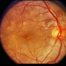
 Fuch's Spot
Apr 2 2019 by Gary R. Cook, MD, FACS
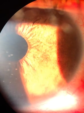
20-year-old patient with high myopia and a Fuch's spot OD.
Condition/keywords: Fuchs, high myopia, pathologic myopia
-
 Fuchs' Heterochromic iridocyclitis
Mar 5 2021 by Niloofar Piri, MD
Heterochromia associated with Fuchs' heterochromic iridocyclitis in the left eye of a 63-year-old patient. Notice the green color of the left iris resulting from diffuse iris atrophy.
Photographer: Douglas Snyder, MD. St. Louis University
Condition/keywords: Fuchs, Fuchs' heterochromic cyclitis, heterochromia
-
 Russell bodies Observed in a Patient with Fuchs' Heterochromic Iridocyclitis
Nov 1 2016 by PAVEL FLORES-MORENO
Anterior chamber shows in iris surface: small, refractile iris crystals.
Photographer: Pavel Flores-Moreno
Imaging device: Anterior chamber camera
Condition/keywords: Fuchs, Russell bodies
-
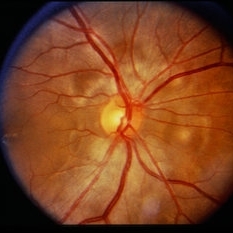
 Slide 2-35
Feb 19 2019 by Lancaster Course in Ophthalmology
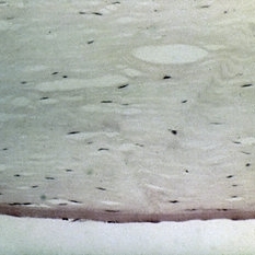
Atrophic iris in Fuch's heterochromic iridocyclitis. Mild plasma cell and lymphocytic infiltrate is present, as well as small vessels in the anterior border layer.
Condition/keywords: Fuchs' heterochromic cyclitis, iris atrophy, lymphocytes
-
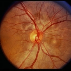
 Slide 7-49
Feb 25 2019 by Lancaster Course in Ophthalmology
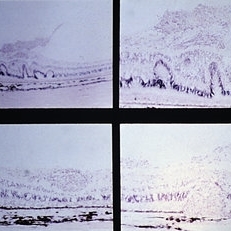
Fuchs' dystrophy is characterized by loss of endothelial cells, thickening of Descemet's membrane, and guttate excrescences of Descemet's membrane.
Condition/keywords: descemet's membrane, endothelial, Fuchs
-
 Slide 9-31
Feb 26 2019 by Lancaster Course in Ophthalmology
Sarcoidosis with retinal and vitreous involvement. A Daten-Fuchs-like nodule of sarcoid granuloma is present (lower right).
Condition/keywords: Dalen-Fuchs nodules, sarcoid granuloma, sarcoidosis
-
 Sunset Glow Fundus
Nov 11 2024 by KANWALJEET HARJOT MADAN, M.S. (Ophthalmology); FAICO (Vitreous - Retina)
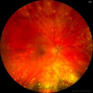
This is fundus picture of right eye of a 15 year-old female patient depicting convalescent phase of Vogt Koyanagi Harada Syndrome. This phase occurs few weeks to few months after the acute stage of the disease. Fundus has a typical bright orange appearance, also known as SUNSET GLOW, along with appearance of DALEN FUCHS nodules.
Photographer: Dr. Kanwaljeet Harjot Madan, M.S. (Ophthalmologist) Fellow in Vitrous & Retina. Thind Eye Hospital, Jalandhar City. Punjab. India
Imaging device: Zeiss Clarus
Condition/keywords: Dalen-Fuchs nodules, Sunset Glow Fundus, VOGT KOYANAGI HARADA
-
 Sympathetic Ophthalmia
Sep 28 2012 by Joseph M. Civantos, MD
59-year-old lady had blunt trauma left eye with a ruptured globe. She refused further surgery on that eye. She returned 4 months later with decreased vision in the right eye. RE dropped from 20/25 to 20/50. The view is hazy due to vitreous cells. Large white choroidal Dalen-Fuchs nodules are visible.
Condition/keywords: Dalen-Fuchs nodules, sympathetic ophthalmia
-
 Vogt-Koyanagi-Harada Disease
Feb 20 2015 by H. Michael Lambert, MD
Color photo showing multifocal detachments of the neurosensory retina with underlying cream colored lesions ( possibly Dalen-Fuchs nodules). Large pocket of subretinal fluid in the macula.
Condition/keywords: exudative detachment, Vogt-Koyanagi-Harada
-
 Vogt-Koyanagi-Harada Disease
Feb 20 2015 by H. Michael Lambert, MD
Color photo showing multifocal detachments of the neurosensory retina with underlying cream colored lesions ( possibly Dalen-Fuchs nodules). Large pocket of subretinal fluid in the macula.
Condition/keywords: exudative retinal detachment, Vogt-Koyanagi-Harada
-
 Vogt-Koyanagi-Harada Disease
Feb 20 2015 by H. Michael Lambert, MD
Dalen-Fuchs' nodule. A granuloma made up of epithelioid cells and lymphocytes between Bruch’s membrane and the retinal pigment epithelium.
Condition/keywords: exudative retinal detachment, Vogt-Koyanagi-Harada
-
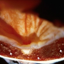 Vogt-Koyanagu-Harada (VKH) Disease
May 18 2020 by McGill University Health Centre
VKH disease is an autoimmune condition that causes bilateral chronic granulomatous panuveitis, and extraocular manifestations in the central nervous system, auditory system, and integument. VKH disease is twice as prevalent in women than men, and is believed to be associated with specific human leukocyte antigen (HLA) types, suggesting a possible hereditary component. The exact cause of VKH disease, however, remains unclear. Image (B) shows several aggregate RPE cells and histiocytes, called Dalen–Fuchs nodules (arrows)
Condition/keywords: Dalen-Fuchs nodules, enucleation, Vogt-Koyanagi-Harada
 Loading…