Search results (8 results)
-
 Fractal Pattern of Chronic Serpiginous Choroiditis
Jun 17 2025 by Guilherme Sturzeneker, MD, MSc
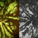
Ultra-widefield fundus photograph and autofluorescence of a 33-year-old woman with longstanding serpiginous choroiditis in the right eye. The image reveals centrifugal chorioretinal atrophy forming a dramatic fractal-like pattern, sparing the fovea. The patient is several years post-onset, with repeated negative workups, including for tuberculosis. Despite extensive lesions, the patient retains 20/20 vision in both eyes. Management included azathioprine monotherapy, as systemic steroids were contraindicated due to bipolar disorder.
Photographer: Andrea Almeida, IPEPO - Instituto da Visão
Imaging device: Optos Silverstone
Condition/keywords: autoimmune uveitis, azathioprine, chorioretinal atrophy, serpiginous choroiditis, ultra-wide field imaging
-
 Susac's Syndrome
Feb 13 2018 by John S. King, MD
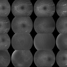
Background: 46-year-old WF with CML (stable on Sprycel) saw her PCP for headaches without known cause; Headaches worsened and became confused, disoriented, off balance, and impaired short term memory. Heme-oncology ordered MRI that showed abnormal signal in the cerebellum and other parts of the brain, and LP has elevated protein. LP did show positive tau test, but fortunately, was a false positive for CJD. IV and PO steroids started and symptoms improved. MRI showed much improvement one month since starting steroids. 3 weeks later had a scotoma in right eye and eye doctor did not find anything at that time to cause it. Tinnitus developed (and some intermittent vertigo before that) and ENT referred back to eye doctor, who then referred the patient to Dr. Zocchi. He found a CWS and superotemporal BRAO OD, and bilateral arteritis (initial FAs). She had some additional work-up for vasculitis. Given the retinal arteritis, cochlear issues, and MRI findings, Dr.Zocchi suspected Susac's Syndrome. She was started on multiple regimens including prednisone, IVIG, azathioprine, and MTX, and has had the best reponse to IVIG. She is stable and doing well with 20/20 vision in both eyes.
Photographer: Maycey Highfill
Imaging device: Topcon
Condition/keywords: Susac's syndrome
-
 Susac's Syndrome
Feb 13 2018 by John S. King, MD
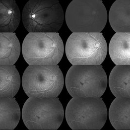
Background: 46-year-old WF with CML (stable on Sprycel) saw her PCP for headaches without known cause; Headaches worsened and became confused, disoriented, off balance, and impaired short term memory. Heme-oncology ordered MRI that showed abnormal signal in the cerebellum and other parts of the brain, and LP has elevated protein. LP did show positive tau test, but fortunately, was a false positive for CJD. IV and PO steroids started and symptoms improved. MRI showed much improvement one month since starting steroids. 3 weeks later had a scotoma in right eye and eye doctor did not find anything at that time to cause it. Tinnitus developed (and some intermittent vertigo before that) and ENT referred back to eye doctor, who then referred the patient to Dr. Zocchi. He found a CWS and BRAO superitemporally OD, and bilateral arteritis (see initial FA). She had some additional work-up for vasculitis. Given the retinal arteritis, cochlear issues, and MRI findings, Dr.Zocchi suspected Susac's Syndrome. She was started on multiple regimens including prednisone, IVIG, azathioprine, and MTX, and has had the best reponse to IVIG. She is stable and doing well with 20/20 vision in both eyes.
Photographer: Maycey Highfill
Imaging device: Topcon
Condition/keywords: Susac's syndrome
-
 Susac's Syndrome
Feb 13 2018 by John S. King, MD
Background: 46-year-old WF with CML (stable on Sprycel) saw her PCP for headaches without known cause; Headaches worsened and became confused, disoriented, off balance, and impaired short term memory. Heme-oncology ordered MRI that showed abnormal signal in the cerebellum and other parts of the brain, and LP has elevated protein. LP did show positive tau test, but fortunately, was a false positive for CJD. IV and PO steroids started and symptoms improved. MRI showed much improvement one month since starting steroids. 3 weeks later had a scotoma in right eye and eye doctor did not find anything at that time to cause it. Tinnitus developed (and some intermittent vertigo before that) and ENT referred back to eye doctor, who then referred the patient to Dr. Zocchi. He found a CWS and BRAO superotemporally OD (see photo), and bilateral arteritis. She had some additional work-up for vasculitis. Given the retinal arteritis, cochlear issues, and MRI findings, Dr.Zocchi suspected Susac's Syndrome. She was started on multiple regimens including prednisone, IVIG, azathioprine, and MTX, and has had the best reponse to IVIG. She is stable and doing well with 20/20 vision in both eyes.
Photographer: Macey Highfill
Imaging device: Topcon
Condition/keywords: Susac's syndrome
-
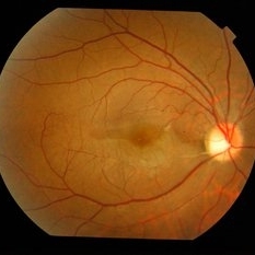
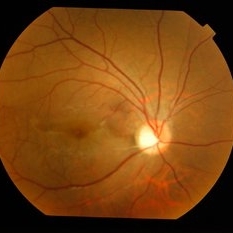
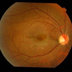 Cilioretinal Artery Occlusion in SLE
May 3 2015 by Mallika Goyal, MD
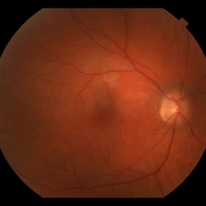
Right eye of a 27 year old lady showing cilioretinal artery occlusion with corresponding macular infarct inferior to centre. She presented with a complaint of a field defect superior to centre, VA was 20/20. She has SLE and is using oral steroids, azathioprine and warfarin (for recent gangrene toes).
Photographer: Mallika Goyal, MD, Apollo Health City, Jubilee Hills, Hyderabad
Condition/keywords: cilioretinal artery occlusion
-
 Cilioretinal Artery Occlusion in SLE
May 3 2015 by Mallika Goyal, MD
Right eye of a 27-year-old lady showing cilioretinal artery occlusion with corresponding macular infarct inferior to centre. She presented with a complaint of a field defect superior to centre, VA was 20/20. She has SLE and is using oral steroids, azathioprine and warfarin (for recent gangrene toes).
Photographer: Mallika Goyal, MD, Apollo Health City, Jubilee Hills, Hyderabad
Condition/keywords: cilioretinal artery occlusion
-
 Cilioretinal artery occlusion in SLE
May 3 2015 by Mallika Goyal, MD
Right eye of a 27-year-old lady showing cilioretinal artery occlusion with corresponding macular infarct inferior to centre. She presented with a complaint of a field defect superior to centre, VA was 20/20. She has SLE and is using oral steroids, azathioprine and warfarin (for recent gangrene toes).
Photographer: Mallika Goyal, MD, Apollo Health City, Jubilee Hills, Hyderabad
Condition/keywords: cilioretinal artery occlusion
 Loading…