Search results (102 results)
-
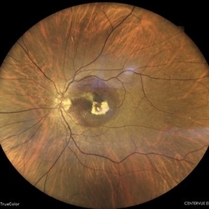
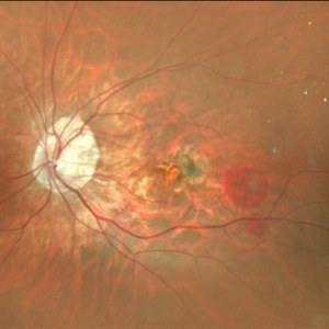 Subretinal Neovascular Membrane
Aug 15 2025 by Akansha Sharma
Color fundus photograph of a 40 year old male with subretinal neovascular membrane.
Photographer: DR. AKANSHA SHARMA
Condition/keywords: choroidal neovascular membrane (CNVM), CNVM, SRNVM, subretinal neovascularization (SRNV), wet age-related macular degeneration (wet AMD)
-
OCT Video Imaging of Left Eye Age Related Macular Degeneration
Jan 6 2025 by Kavitha Duraipandi, MD DNB FICO FRCS
Left eye OCT macula shows various biomarkers like PED, sub retinal fluid, sub retinal hyper reflective material and hyper reflective foci suggestive of wet age-related macular degeneration.
Condition/keywords: OCT biomarkers, wet age-related macular degeneration (wet AMD)
-
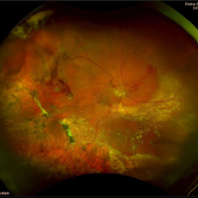 Peripheral Exudative Hemorrhagic Chorioretinopathy
Nov 19 2024 by Toolie Winters
Ultra-wide field fundus photograph of an 85-year-old woman with Peripheral Exudative Hemorrhagic Chorioretinopathy (PECHR) affecting the right eye. Patient presented with a blind spot centrally in the right eye which she first noticed 4 months prior to this image being taken. The patient states that in the month prior to this image, she began noticing bright lights flash across her vision 4-5x/day which last about 15 seconds. The flashes are either black with a blue ring around them or yellow, and their frequency has increased over time. The patient's vision at the time of this appointment was Dcc20/100+1 PHNI. This photo also shows diffuse hemorrhage, lipid, and an eccentric disciform lesion.
Photographer: Toolie Winters
Imaging device: Optos California
Condition/keywords: fundus photography, neovascular age-related macular degeneration (AMD), Optos, OPTOS CALIFORNIA, peripheral exudative hemorrhagic chorioretinopathy (PEHCR), pseudocolor, ultra-wide field imaging, wet age-related macular degeneration (wet AMD)
-
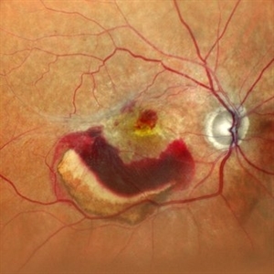
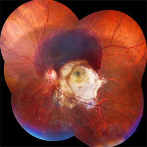
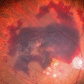 Large Subretinal Bleed in Case of Wet ARMD
Sep 28 2024 by Anjana Mirajkar, MS Ophthalmology
An intra operative image showing large sub retinal hemorrhage involving the macular area and along the superior arcade with exudation at the macular area in case of wet ARMD.
Photographer: Dr. Anjana Mirajkar -Retina Foundation, Ahmedabad
Condition/keywords: polypoidal choroidal vasculopathy (PCV), subretinal hemorrhage, wet age-related macular degeneration (wet AMD)
-
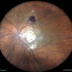
 Large PED with RPE-rip
Aug 21 2024 by Amirfarbod Yazdanyar, MD , PhD
Fundus photograph of a 85 year-old male with AMD who developed very large PED with RPE-rip. BCVA is 20/70
Photographer: Justin Cocilo , Retina Group of New England
Condition/keywords: large pigment epithelial detachment, RPE-rip, wet age-related macular degeneration (wet AMD)
-
 RPE-Transplantation
Jul 25 2024 by Gabriel Costa Andrade, PhD
Postoperative period of RPE-transplantation in a patient with neovascular AMD after RPE tear.
Photographer: Gabriel Andrade
Condition/keywords: neovascular age-related macular degeneration (AMD), pars plana vitrectomy (PPV), wet age-related macular degeneration (wet AMD)
-
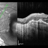
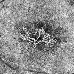
 Neovascular AMD with Polypoidal Choroidal Vasculopathy
Jun 11 2024 by Gregg T. Kokame, MD, MMM, FASRS
Structure OCT from the OCT angiography shows definition of the PCV complex.
Photographer: Jaclyn Pisano
Condition/keywords: branching vascular network (BVN), OCTA, polypoidal choroidal vasculopathy (PCV), wet age-related macular degeneration (wet AMD)
-
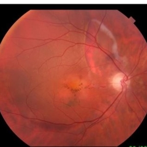
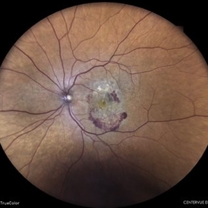
 Subretinal Neovascular Membrane
Jun 5 2024 by Akansha Sharma
Color fundus photograph of a 49 year old female with subretinal bleed suggestive of subretinal neovascular membrane.
Photographer: Dr. Akansha Sharma, Bharati Eye Hospital
Condition/keywords: choroidal neovascular membrane (CNVM), CNVM, SRNVM, subretinal neovascularization (SRNV), wet age-related macular degeneration (wet AMD)
-
 Subretinal Neovascular Membrane
Jun 5 2024 by Akansha Sharma
Color fundus photograph of a 94 year old female with subretinal bleed suggestive of subretinal neovascular membrane.
Photographer: Dr. Akansha Sharma, Bharati Eye Hospital
Condition/keywords: choroidal neovascular membrane (CNVM), CNVM, SRNVM, subretinal neovascularization (SRNV), wet age-related macular degeneration (wet AMD)
-
 Subretinal Neovascular Membrane with PED
Apr 17 2024 by Akansha Sharma
Color fundus photograph of a 72 year old male with ped along with subretinal bleed around it.
Photographer: Dr. Akansha Sharma, Bharati Eye Hospital
Condition/keywords: CNVM, PED, SRNVM, subretinal neovascularization (SRNV), wet age-related macular degeneration (wet AMD)
-
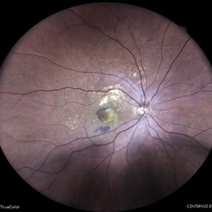
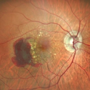
 Idiopathic Polypoidal Choroidal Vasculopathy
Apr 9 2024 by Akansha Sharma
Color fundus photograph of a 74 year old female with subretinal bleed in a case of polypoidal choroidal vasculopathy.
Photographer: Dr. Akansha Sharma, Bharati Eye Hospital
Condition/keywords: PCV, polypoidal choroidal vasculopathy (PCV), wet age-related macular degeneration (wet AMD)
-
 Subretinal Neovascular Membrane
Mar 26 2024 by Akansha Sharma
Color fundus photograph of a 65 year old female patient with subretinal bleed suggestive of subretinal neovascular membrane.
Photographer: Dr. Akansha Sharma, Bharati Eye Hospital
Condition/keywords: CNVM, SRNVM, subretinal neovascularization (SRNV), wet age-related macular degeneration (wet AMD)
-
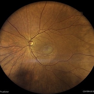
 Ghost of Halloween
Mar 22 2024 by Tushar Agrawal
Fundus photograph of a 57-year-old man with a macular choroidal neovascular membrane observed over time.
Condition/keywords: choroidal neovascular membrane (CNVM), macula choroidal neovascularization, wet age-related macular degeneration (wet AMD)
-
 Wet age related macular degeneration
Jan 28 2024 by Anjana Mirajkar, MS Ophthalmology
Fundus photograph of an 70 year old male with sub retinal bleed and exudation as well as scarring in case of wet age related macular degeneration.
Photographer: Dr. Anjana Mirajkar -Retina Foundation, Ahmedabad
Imaging device: Mirante-Nidek
Condition/keywords: choroidal neovascular membrane (CNVM), wet age-related macular degeneration (wet AMD)
-
 Neovascular-network-OCTA
Jan 2 2024 by Tahsin Khundkar, MD
En Face optical coherence tomography (OCT).- angiography shows a large choroidal neovascular membrane in the outer retina to choriocapillaris slab.
Photographer: Jeffrey Zeigler, Concord Eye Center
Imaging device: Zeiss
Condition/keywords: neovascular membrane, wet age-related macular degeneration (wet AMD)
-
 Neovascular AMD with Ring Shaped lesions
Jul 12 2023 by Gregg T. Kokame, MD, MMM, FASRS
Horizontal OCT Scan - Neovascular AMD with Active CNV Ring shaped lesions underneath the RPE inverted U-shaped elevation
Photographer: Jaclyn Pisano
Imaging device: Zeiss Cirrus 6000
Condition/keywords: inverted u-shaped elevation, macular edema, OCT, Sub-retinal fluid, wet age-related macular degeneration (wet AMD)
-
 Neovascular AMD with Ring Shaped lesions
Jul 12 2023 by Gregg T. Kokame, MD, MMM, FASRS
Vertical OCT Scan - Neovascular AMD with Active CNV Ring shaped lesions underneath the RPE inverted U-shaped elevation
Photographer: Jaclyn Pisano
Imaging device: Zeiss Cirrus 6000
Condition/keywords: edema, OCT, Sub-retinal fluid, wet age-related macular degeneration (wet AMD)
-
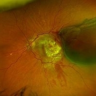
 SCARRED CHOROIDAL NEOVACULAR MEMBRANE
Jun 6 2023 by Akansha Sharma
COLOUR FUNDUS PHOTOGRAPH OF A 82 YEAR OLD MALE WITH SCARRED CHOROIDAL NEOVASCULAR MEMBRANE
Photographer: Dr. Urmil Shah, Dr. Akansha Sharma, Dr. Denish Patel
Condition/keywords: choroidal neovascular membrane (CNVM), wet age-related macular degeneration (wet AMD)
-
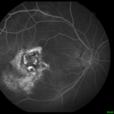
 Lady in a dress
Feb 9 2023 by Shelby Helton
Fluorescein Angiography on a 67-year-old male with significant RPE changes secondary to a severe subretinal hemorrhage that required a vitrectomy with subretinal TPA in 2013.
Photographer: Shelby Helton
Imaging device: Heidelberg Spectralis
Condition/keywords: wet age-related macular degeneration (wet AMD)
-
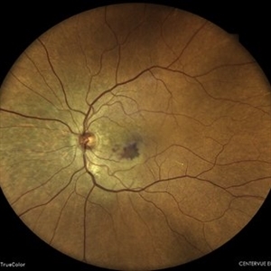
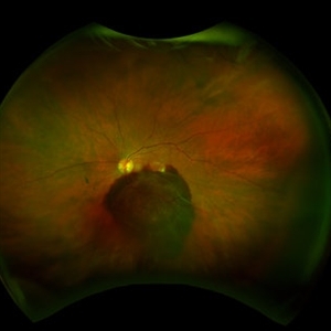
 WET AGE RELATED MACULAR DEGENERATION
Nov 21 2022 by Akansha Sharma
COLOUR FUNDUS PHOTOGRAPH OF A 71 YEAR OLD MALE WITH SUBRETINAL BLEED IN A CASE OF WET AGE RELATED MACULAR DEGENERATION
Photographer: Dr. Akansha Sharma-Retina Foundation, Ahmedabad
Condition/keywords: CNVM, subretinal neovascularization (SRNV), wet age-related macular degeneration (wet AMD)
-
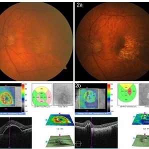 Pigment-epitelial-tear-in-AMD
Mar 20 2022 by Argyrios Chronopoulos, MD, FMH, MRCP
Image 1a Fundus image of a wet AMD with PED (OCT image 1b), Image 2a RPE tear following anti-VEGF treatment (OCT image 2b)
Photographer: A. Chronopoulos, Ludwigshafen Hopsital
Imaging device: Fundus imaging device
Condition/keywords: antiVEGF therapy, wet age-related macular degeneration (wet AMD)
-
 Subretinal Hemorrhage and Disciform Scar
Feb 1 2022 by Lucas Zago Ribeiro, MD
Fundus photo of a 70-year-old man with acute subretinal hemorrhage, and previous disciform scar due to Wet AMD.
Photographer: Lucas Zago Ribeiro, UNIFESP, Brazil
Condition/keywords: disciform scar, subretinal hemorrhage, wet age-related macular degeneration (wet AMD)
-
 Hemorrhagic Choroidal Detachment
Jul 6 2021 by Kristen Wagner
Hemorrhagic choroidal detachment with overlying retinal detachment, neovascular AMD with inactive scar.
Photographer: Kristen Wagner COT, Tennessee Retina, Nashville TN
Imaging device: optos
Condition/keywords: choroidal detachment, hemorrhage, wet age-related macular degeneration (wet AMD)
-
 Choroidal Neovascular Membrane Evolving With Subretinal Hemorrhage
Apr 23 2021 by Andre Beckenkamp
Wide angle fundus photograph of an 82-year-old woman with dry AMD in her right eye and wet AMD in the left eye, evolving with subretinal hemorrhage and associated serous retinal detachment.
Photographer: Andre Beckenkamp
Imaging device: Optos Daytona
Condition/keywords: age-related macular degeneration (AMD), serous retinal detachment, subretinal hemorrhage, wet age-related macular degeneration (wet AMD)
-
 Management of Submacular Hemorrhage
Mar 22 2020 by Anfisa Ayalon, MD
Intraoperative images were taken during the management of submacular hemorrhage in age-related macular degeneration. The goal of the surgery was the physical displacement of SMH out of the fovea using expansile gas. The image from the left was done during ILM peeling. Note the massive collection of subretinal blood. The image from the right was done after submacular injection of t-PA, bevacizumab and filtered air. Intravitreal injection of 20% SF6 completed the surgery. The visual acuity improved after the surgery from HM to 1/60.
Photographer: Anfisa Ayalon, MD., Meir Medical Center, Kfar Saba, Israel.
Condition/keywords: spontaneous submacular hemorrhage, submacular hemorrhage, vitreomacular surgery, wet age-related macular degeneration (wet AMD)
 Loading…