Search results (43 results)
-
 Adult Onset Vitelliform Macular Dystrophy
Sep 29 2024 by Tejaswita Verma
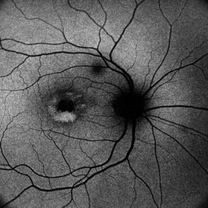
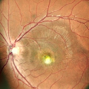
Autofluorescence image of the RE of a 62 year-old hypertensive female with 6/12 vision showing hyperautofluorescence . The patient was asked to review every few months to check for development of secondary CNVM
Photographer: DR. TEJASWITA VERMA
Imaging device: MIRANTE
Condition/keywords: Adult vitelliform macular dystrophy
-
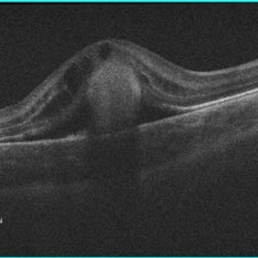
 OCT in Adult Vitelliform Dystrophy
Jun 25 2024 by Tejaswita Verma
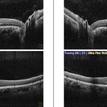
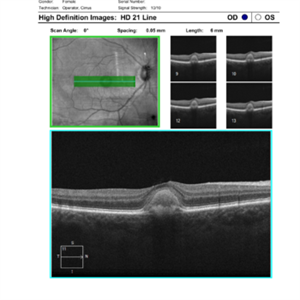
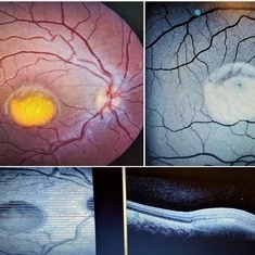
OCT image of a 62 year old female with 6/12 vision in both eyes showing sub retinal fluid with RPE granularity s/o Adult vitelliform macular dystrophy.
Photographer: DR. TEJASWITA VERMA
Imaging device: MIRANTE
Condition/keywords: adult vitelliform dystrophy, optical coherence tomography (OCT)
-
 Adult Vitelliform Macular Dystrophy
Jun 25 2024 by Tejaswita Verma
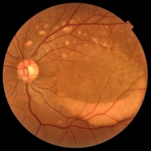
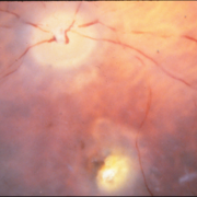
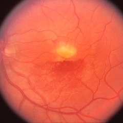
Right eye Fundus photo of an elderly 62 year old female with 6/12 vision showing elevated lesion at macula with suggestive of adult vitelliform macular dystrophy, misdiagnosed as long standing CSR elsewhere.
Photographer: DR. TEJASWITA VERMA
Imaging device: MIRANTE
Condition/keywords: Adult-onset vitelliform dystrophy
-
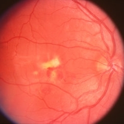
 Adult Vitelliform Macular Dystrophy
Jun 25 2024 by Tejaswita Verma
Left eye fundus photograph of an elderly 62 year old hypertensive female showing elevated lesion at macula s/o adult vitelliform macular dystrophy, misdiagnosed as long standing CSR elsewhere.
Photographer: DR. TEJASWITA VERMA
Imaging device: MIRANTE
Condition/keywords: Adult vitelliform macular dystrophy
-
 Representative Electrooculogram Responses
May 13 2024 by Gabrielle Hallai
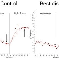
Electrooculogram responses on the left from a control individual with no known retinal pathology. There is a clear dark trough around 13 minutes (arrow down) and a light peak around 25 minutes (arrow up). The Arden ratio, or the light peak to dark trough ratio, is 2.54, indicative of normal retinal pigment epithelium function (normal > 1.80, abnormal < 1.65). On the right-hand side, there is a representative image from an individual with Best macular dystrophy. Note the reduced responses for both the dark and light phase. There is a reduced Arden ratio of 1.23, suggestive of abnormal retinal pigment epithelium function. An abnormal Arden ratio is universal in Best vitelliform macular dystrophy and is the most common electroretinographic change in this disease. Other bestrophinopathies such as autosomal recessive bestrophinopathy may have normal EOG. EOG testing was completed on the Diagnosys ColorDome.
Photographer: Gabrielle Hallai, PhD, Cleveland Clinic Cole Eye Institute
Imaging device: Diagnosys ColorDome
Condition/keywords: Best disease, electrooculogram, electroretinography, EOG
-
 Best Disease
Apr 24 2024 by Marcelo Zas, MD PhD
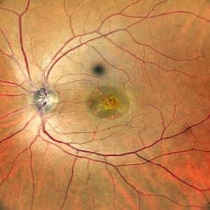
Best vitelliform macular dystrophy (BVMD) or Best disease. Is the most common autosomal dominant macular dystrophy. It involves the retinal pigment epithelium (RPE), and leads to a characteristic bilateral yellow “egg-yolk” appearance of the macula as you can see in this image. Essentially, BVMD is considered to have 6 clinical stages: Previtelliform, Vitelliform, Pseudohypopyon, Vitelleruptive, Atrophic and Choroidal neovascularization. As the disease progresses, patients may experience a slow, bilateral decrease in visual acuity, central scotoma, or metamorphopsia. With secondary CNV, visual decline can be rapid, however.
Photographer: Luciano Scorsetti MD
Condition/keywords: Macular Dystrophy
-
 Multifocal Best Disease
Feb 6 2024 by KRISHNENDU NANDI, MS
A 38-year-old male presented with gradual dimness of vision in both eyes for last 3 months. Best corrected visual acuity was 6/24, N8 in both eyes. Colour fundus photograph showed multiple orangish yellow sub retinal lesions on the posterior pole extending beyond arcades. Macular thickening also noted. OCT line scan through the fovea showed thickened ellipsoid zone and it was separated from the RPE by optically clear space.
Photographer: Dr. Krishnendu Nandi
Condition/keywords: Best vitelliform macular dystrophy (BVMD), Multifocal
-
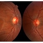
 Best Vitelliform Macular Dystrophy
Jun 26 2022 by Vaidehi Sathaye
8 yr old male child with Best Vitelliform Macular Dystrophy
Photographer: Dr. Vaidehi Sathaye
Imaging device: Mirante
Condition/keywords: Best vitelliform macular dystrophy (BVMD)
-
 Best Vitelliform Macular Dystrophy
Jun 26 2022 by Vaidehi Sathaye
8 yr old male child with Best Vitelliform Macular Dystrophy
Photographer: Dr. Vaidehi Sathaye
Imaging device: Mirante
Condition/keywords: Best vitelliform macular dystrophy (BVMD)
-
 Adult-onset foveomacular vitelliform dystrophy
May 26 2022 by Rinat Sutiushev
Patient born in 1946. Concerns about decreased vision in right eye, distortions when reading. The ocular fundus of both eyes shows round yellowish deposits (vitelliform material deposits) in the fovea. Autofluorescence photography reveals hyperautofluorescence. OCT demonstrates the presence of vitelliform material under the sensitive retina and over the retinal pigment epithelium.
Photographer: Rinat Sutiushev
Condition/keywords: adult vitelliform dystrophy, vitelliform lesion, vitelliform macular dystrophy
-
 Adult-onset foveomacular vitelliform dystrophy
May 26 2022 by Rinat Sutiushev
Woman born in 1946. Concerns about decreased vision in right eye, distortions when reading. The ocular fundus of both eyes shows round yellowish deposits (vitelliform material deposits) in the fovea. When autofluorescence photography is performed, hyperautofluorescence is detected.
Photographer: Rinat Sutiushev
Condition/keywords: adult vitelliform dystrophy, vitelliform lesion, vitelliform macular dystrophy
-
 Adult-onset foveomacular vitelliform dystrophy
May 26 2022 by Rinat Sutiushev
Woman born in 1946. Concerns about decreased vision in the right eye, distortions when reading. The ocular fundus of both eyes shows round yellowish deposits (vitelliform material deposits) in the fovea.
Photographer: Rinat Sutiushev
Condition/keywords: adult vitelliform dystrophy, vitelliform lesion, vitelliform macular dystrophy
-
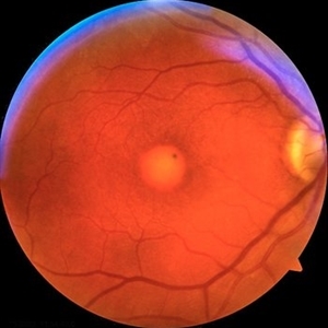
 Best Vitelliform Macular Dystrophy
Dec 10 2020 by McGill University Health Centre
Postmortem eyes from 101-year-old female. Past clinical history includes a poor vision for many years due to macular degeneration. The last visual acuity test recorded 6/15 OD and 6/6 OS. IOP 14 and 18 torr OS. Histopathology: Disclosed and yellow 2x2mm macular lesion. Microscopic examination: elevated placoid macular lesion with overlying pigment granules. Electron microscopy examination: pigment granules with abundant lipofuscin and melanolysosomes, photoreceptor cells markedly attenuated (less degenerated at the periphery) Numerous calcified drusen throughout the retina particularly in the posterior pole. RPE lipofuscin content is elevated in Best’s dystrophy. The extractability of the PRE lipofuscin fluorophores is reduced (it is normal during senescence). The defect in Best’s dystrophy accelerates this age related change in lipofuscin.
Condition/keywords: Best vitelliform macular dystrophy (BVMD), histopathology, pathology
-
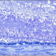
 Best Vitelliform Macular Dystrophy
Dec 10 2020 by McGill University Health Centre
Postmortem eyes from 101-year-old female. Past clinical history includes a poor vision for many years due to macular degeneration. The last visual acuity test recorded 6/15 OD and 6/6 OS. IOP 14 and 18 torr OS. Histopathology: Disclosed and yellow 2x2mm macular lesion. Microscopic examination: elevated placoid macular lesion with overlying pigment granules. Electron microscopy examination: pigment granules with abundant lipofuscin and melanolysosomes, photoreceptor cells markedly attenuated (less degenerated at the periphery) Numerous calcified drusen throughout the retina particularly in the posterior pole. RPE lipofuscin content is elevated in Best’s dystrophy. The extractability of the PRE lipofuscin fluorophores is reduced (it is normal during senescence). The defect in Best’s dystrophy accelerates this age related change in lipofuscin.
Condition/keywords: Best vitelliform macular dystrophy (BVMD), histopathology, pathology
-
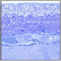
 Best Vitelliform Macular Dystrophy
Dec 10 2020 by McGill University Health Centre
Postmortem eyes from 101-year-old female. Past clinical history includes a poor vision for many years due to macular degeneration. The last Visual acuity test recorded 6/15 OD and 6/6 OS. IOP 14 and 18 torr OS. Histopathology: Disclosed and yellow 2x2mm macular lesion. Microscopic examination: elevated placoid macular lesion with overlying pigment granules. Electron microscopy examination: pigment granules with abundant lipofuscin and melanolysosomes, photoreceptor cells markedly attenuated (less degenerated at the periphery) Numerous calcified drusen throughout the retina particularly in the posterior pole. RPE lipofuscin content is elevated in Best’s dystrophy. The extractability of the PRE lipofuscin fluorophores is reduced (it is normal during senescence). The defect in Best’s dystrophy accelerates this age related change in lipofuscin.
Condition/keywords: Best vitelliform macular dystrophy (BVMD), histopathology, pathology
-
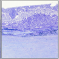
 Best Vitelliform Macular Dystrophy
Dec 10 2020 by McGill University Health Centre
Postmortem eyes from 101-year-old female. Past clinical history includes a poor vision for many years due to macular degeneration. The last visual acuity test recorded 6/15 OD and 6/6 OS. IOP 14 and 18 torr OS. Histopathology: Disclosed and yellow 2x2mm macular lesion. Microscopic examination: elevated placoid macular lesion with overlying pigment granules. Electron microscopy examination: pigment granules with abundant lipofuscin and melanolysosomes, photoreceptor cells markedly attenuated (less degenerated at the periphery) Numerous calcified drusen throughout the retina particularly in the posterior pole. RPE lipofuscin content is elevated in Best’s dystrophy. The extractability of the PRE lipofuscin fluorophores is reduced (it is normal during senescence). The defect in Best’s dystrophy accelerates this age related change in lipofuscin.
Condition/keywords: Best vitelliform macular dystrophy (BVMD), fundus photograph
-
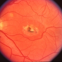
 Best Vitelliform Macular Dystrophy
Mar 17 2020 by Sophia El Hamichi, MD
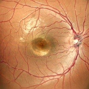
Classic "egg yolk" presentation in a 16-year-old female with best disease.
Condition/keywords: autofluorescence imaging, Best disease, optical coherence tomography (OCT), vitelliform macular dystrophy
-
 Best's Vitelliform Macular Dystrophy
Apr 8 2019 by Gary R. Cook, MD, FACS
Left eye of 29-year-old white male with Best's disease showing only a little yellow material and more pigmentation of the lesion; VA = 20/200
Condition/keywords: Best disease, vitelliform macular dystrophy
-
 Best's Vitelliform Macular Dystrophy
Apr 8 2019 by Gary R. Cook, MD, FACS
Right eye of a 29-year-old white male with Best's disease; OD; VA = 20/25.
Condition/keywords: Best disease, vitelliform macular dystrophy
-
 Best Disease
Apr 8 2019 by Gary R. Cook, MD, FACS
Left eye of the patient with Best disease and an active CNV OD showing resolving hemorrhage from prior CNV OS.
Condition/keywords: Best disease, retinal hemorrhage, vitelliform macular dystrophy
-
 Best Disease
Apr 8 2019 by Gary R. Cook, MD, FACS
Right eye of patient with Best disease with active CNV OD; V.A. = 20/30.
Condition/keywords: Best disease, choroidal neovascularization (CNV), vitelliform macular dystrophy
-
 Best's Vitelliform Macular Dystrophy
Apr 8 2019 by Gary R. Cook, MD, FACS
Vitelliform lesion in the left eye of a 10-year-old female demonstrating a yellow, round, egg yolk-like lesion; V. A. = 20/25
Condition/keywords: Best disease, vitelliform lesion, vitelliform macular dystrophy
-
 Best Vitelliform Dystrophy with Secondary CNVM
Jan 8 2019 by Rutul R Patel, MD Ophthalmology
10-year-old girl with b/l Best vitelliform dystrophy and left eye secondary CNVM.
Photographer: Dr. Rutul Patel
Imaging device: TOPCON TRC 50DX
Condition/keywords: Best disease, choroidal neovascular membrane (CNVM), vitelliform macular dystrophy
-
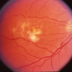
 Best Disease
May 1 2018 by Mitzy E Torres Soriano, MD
Fundus photographs of an 5-year-old boy with best vitelliform macular dystrophy and family history.
Photographer: Luciana García,MD
Condition/keywords: Best disease, vitelliform macular dystrophy
-
 Cystoid Macular Edema (CME) in Vitelliform Macular Dystrophy (VMD)
Apr 22 2018 by Ronald Silva
Macula OCT of a 3-year-old boy with low vision and cystoid macular edema (CME) in vitelliform macular dystrophy (VMD) in right eye.
Photographer: Ronald Rocha da Silva, HCOE, Feira de Santana-BA
Condition/keywords: Best disease, cystoid macular edema (CME), vitelliform macular dystrophy
 Loading…