Search results (187 results)
-
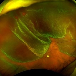
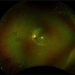 Chorioretinal Macula Scar (Ultrawide View)
May 12 2025 by Briana Hernandez
Ultra wide Optos image of Chorioretinal Macular Scar in 9-year-old female patient.
Photographer: Briana Hernandez, Hilton Head Retina Institute
Imaging device: Optos
Condition/keywords: chorioretinal scar, macular scar, ultra-wide field imaging
-
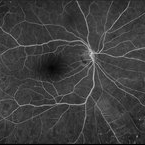 Retinal Vasculitis
Mar 26 2025 by Korey Starkey
41 year-old patient presents with vascular FA findings of occlusive vasculitis with four quadrant Kyrieleis plaques OU showcases a possibly rare but reported atypical presentation of Behcet's Syndrome.
Photographer: Korey Starkey
Imaging device: Optos
Condition/keywords: FA early phase, Fundus Fluorescein Angiography, ischemia, Optos, retinal vasculitis, ultra-wide field imaging, venous beading
-
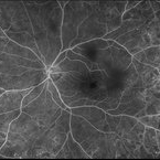 Retinal Vasculitis
Mar 26 2025 by Korey Starkey
41 year-old patient presents with vascular FA findings of occlusive vasculitis with four quadrant Kyrieleis plaques OU showcases a possibly rare but reported atypical presentation of Behcet's Syndrome.
Photographer: Korey Starkey
Imaging device: Optos
Condition/keywords: Behcet's Disease, FA early phase, Fundus Fluorescein Angiography, Optos, retinal vasculitis, ultra-wide field imaging, venous beading
-
 CHRPE
CHRPE
Mar 25 2025 by Toolie Winters
Ultra-wide field fundus photograph of a 78-year-old woman with extensive CHRPE lesions OS. Continued observation has been recommended at this time.
Photographer: Toolie Winters
Imaging device: Optos California
Condition/keywords: CHRPE, congenital hypertrophy of the retinal pigment epithelium (CHRPE), fundus photography, Optos, Optos California, pseudocolor, ultra-wide field imaging
-
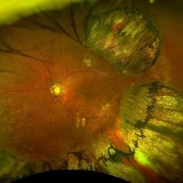
 Retinal Detachment and Lattice Degeneration
Mar 25 2025 by Korey Starkey
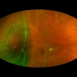
26 year-old patient presented at first visit with rhegmatogenous macula involving retinal detachment of the left eye. Underwent prompt surgical repair. Both eyes also present with lattice degeneration with atrophic holes.
Photographer: Korey Starkey
Condition/keywords: atrophic retinal hole, fundus photography, lattice degeneration, montage photo, Optos, OPTOS CALIFORNIA RGB, retinal detachment, retinal holes, rhegmatogenous retinal detachment, ultra-wide field imaging
-
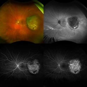 Melanoma Multimodal Evaluation
Feb 10 2025 by Gustavo M. Hüning, MD, MBA, FASRS
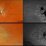
UWF multimodal imaging of an 37-year-old woman with a choroidal melanoma. The mosaic shows a colored retinography; a FAF with regions of previous serous detachments; an early stage of angiography and a later time.
Photographer: Gustavo M. Hüning, HÜNING Clínica do Olhar, Santa Maria - Brazil
Imaging device: Optos California
Condition/keywords: Autofluorescence, Choroidal, Fluorescein angiography, melanoma, multimodal imaging, ultra-wide field imaging
-
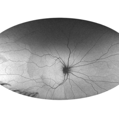 Foveal Hypoplasia AF
Feb 1 2025 by Poornachandra B, MS, FVRS
This is a wide field autofluorescence image of 21 year-old male. He presented with history of low vision since childhood associated with nystagmus. Uniform fluorescence across posterior pole with absent foveal hypo autofluorescence can be seen on the image.
Photographer: Mr Dhikshith
Condition/keywords: autofluorescence imaging, foveal hypoplasia, nystagmus, ultra-wide field imaging
-
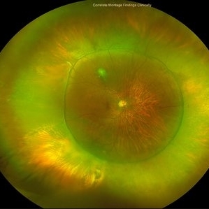
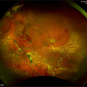 Peripheral Exudative Hemorrhagic Chorioretinopathy
Nov 19 2024 by Toolie Winters
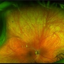
Ultra-wide field fundus photograph of an 85-year-old woman with Peripheral Exudative Hemorrhagic Chorioretinopathy (PECHR) affecting the right eye. Patient presented with a blind spot centrally in the right eye which she first noticed 4 months prior to this image being taken. The patient states that in the month prior to this image, she began noticing bright lights flash across her vision 4-5x/day which last about 15 seconds. The flashes are either black with a blue ring around them or yellow, and their frequency has increased over time. The patient's vision at the time of this appointment was Dcc20/100+1 PHNI. This photo also shows diffuse hemorrhage, lipid, and an eccentric disciform lesion.
Photographer: Toolie Winters
Imaging device: Optos California
Condition/keywords: fundus photography, neovascular age-related macular degeneration (AMD), Optos, OPTOS CALIFORNIA, peripheral exudative hemorrhagic chorioretinopathy (PEHCR), pseudocolor, ultra-wide field imaging, wet age-related macular degeneration (wet AMD)
-
 Occlusive Retinal Vasculitis
Oct 3 2024 by Logan ryzenga
4 view ultra-widefield Optos fluorescein angiogram in the left eye of a 39 year old woman occlusive retinal vasculitis with four quadrant Kyrieleis plaques. This is a showcase of a suspected, rarely reported, and atypical presentation of Behcet's Syndrome.
Photographer: Logan Ryzenga
Imaging device: Optos California
Condition/keywords: Behcet's Disease, Behcet's uveitis, Fluorescein angiography, fluorescein leakage, kyrieleis plaques, non-perfusion, OPTOS, OPTOS CALIFORNIA, ultra-wide field imaging, Uveitis
-
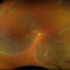 Rhegmatogenous Macula-Off Retinal Detachment
Sep 19 2024 by Alexis Singstock
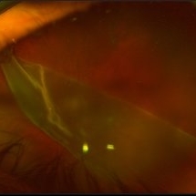
Ultra wide field fundus photograph of a 62 year old female with a rhegmatogenous macula-off retinal detachment affecting her right eye. Patient reported decreased vision, curtain/veil in vision and eye pain with the onset approximately 2 weeks prior to initial encounter.
Photographer: Alexis Singstock
Imaging device: Optos California RGB
Condition/keywords: macula off retinal detachment, Optos, OPTOS CALIFORNIA, OPTOS CALIFORNIA RGB, Retina detachment, ultra-wide field imaging
-
 Stargardt Disease
Aug 27 2024 by Korey Starkey
Ultra wide-field fundus photograph and fundus autofluorescence of a 49-year-old male. Initial visit imaging.
Photographer: Korey Starkey
Imaging device: Optos
Condition/keywords: fundus autofluorescence (FAF), fundus photograph, Optos, Stargardt disease, ultra-wide field imaging
-
 From Ora to Ora
Aug 26 2024 by Nassim Alejandro Abreu Arbaje, MD
Ultra-wide field OCT angiography of a 39 year-old healthy male. The photo attempts to explore retinal vasculature up to the ora serrata.
Photographer: Johel Arrieta, TowardPi
Imaging device: TowardPi BMizar 400khz
Condition/keywords: OCT Angiography, OCTA, ultra-wide field imaging
-
 Rhegmatogenous Macula-On Retinal Detachment (Honeycomb)
Aug 6 2024 by Xitlali Caterina
Ultra-wide field fundus photograph of a 72 year old female with a macula-on retinal detachment with multiple breaks affecting her right eye. Patient presented in the office with flashes of light for five consecutive days prior. The patients vision was sc20/30 PHNI. The physician also noted an acute posterior vitreous detachment and lattice degeneration in the affect eye.
Photographer: Xitlali Caterina
Imaging device: Optos California RGB
Condition/keywords: honeycomb, lattice degeneration, Optos, posterior vitreous detachment, Retinal Detachment with Multiple Breaks, rhegmatogenous retinal detachment, ultra-wide field imaging
-
 Active Proliferative Diabetic Retinopathy
Jul 12 2024 by Korey Starkey
Fluorescein angiogram performed on 35 year old female with active proliferative diabetic retinopathy. Patient has peripapillary vascular loop and history of PRP treatment in both eyes. Patients left eye vision measured at Dcc20/200-1 at this visit.
Photographer: Korey Starkey
Imaging device: Optos
Condition/keywords: FLUORESCEIN ANGIOGRAPHY, hyperfluorescence, laser scarring, Optos, proliferative diabetic retinopathy (PDR), sea fan, ultra-wide field imaging, vascular loop
-
 Panuveitis
Jul 12 2024 by Korey Starkey
Ultra widefield Optos FA of 59 year old female presents with panuveitis in both eyes. Patients vision was VA OS: Dcc20/60-2 at time of visit.
Photographer: Korey Starkey
Imaging device: Optos
Condition/keywords: FLUORESCEIN ANGIOGRAPHY, hyperfluorescence, Optos, Panuveitis, ultra-wide field imaging, Uveitis
-
 Panuveitis
Apr 2 2024 by Zach Seim
Optos Ultra-widefield photo OS of a 59 year old female with Panuveitis OU.
Photographer: Zach Seim
Imaging device: Optos California
Condition/keywords: Optos, OPTOS CALIFORNIA, panuveitis, ULTRA WIDE FIELD, ultra-wide field imaging
-
 Choroidal Melanoma
Mar 26 2024 by Xitlali Caterina
Ultra-widefield fundus photograph of a 40-year-old woman with Choroidal Melanoma in right eye. Patient present with 20/50+2 vision in the right eye. Patient reported having frequent headaches located frontal area of their head and sometimes radiated to the right side as well. Patient also noted eye pain in both eyes that has remained constant for many years, as well as light sensitivity. The physician stated that since this is a medium-sized tumor, the treatment options include I-125 brachytherapy or enucleation. He recommended I-125 brachytherapy.
Photographer: Xitlali Caterina
Imaging device: Optos California RGB
Condition/keywords: fundus photography, Optos, OPTOS CALIFORNIA, superior retina, ultra-wide field imaging, ultra-widefield image
-
 Retinal Detachment with Giant Retinal Tear
Mar 26 2024 by Xitlali Caterina
Ultra-widefield fundus photograph of a 43-year-old male with a Retinal Detachment with Giant Retinal Tear affecting his left eye. Patient presented to the office with count fingers vision at 2 feet. He stated that about 8-9 days ago, he developed a clear curtain/veil and his vision started to get blurry. He also noted that he had floaters and flashes for about 8-9 days as well. The patient had cataract surgery a month prior to his visit. He stated that since his surgery, his vision had been better, but he had an area where he was not able to see well. The physician recommended a complex retinal detachment repair.
Photographer: Xitlali Caterina
Imaging device: OPTOS California RGB
Condition/keywords: fundus photograph, giant retinal tear, left eye, Optos, OPTOS CALIFORNIA, retinal detachment of the macula, retinal detachment with tear, ultra-wide field imaging, ultra-widefield image
-
 Serous Retinal Detachment in Advanced Proliferative Diabetic Retinopathy
Feb 15 2024 by Annaka Gooding
Ultra-Wide fundus photograph of a 29 year old female with a Serous Retinal Detachment in Advanced PDR. Patient present to clinic with LP vision following PPV and fill in PRP. Physician recommended oral prednisone treatment and to reassess at their following visit.
Photographer: Annaka Gooding, CPO
Imaging device: Optos California RGB
Condition/keywords: Diabetes, diabetic macular edema, fundus photography, OPTOS CALIFORNIA, pan-retinal photocoagulation (PRP), pars plana vitrectomy (PPV), proliferative diabetic retinopathy (PDR), serous retinal detachment, ultra-wide field imaging
-
 Macula off Retinal Detachment
Jan 23 2024 by Annaka Gooding
Ultra-widefield fundus photograph of an 81-year-old male with a Macula Off Retinal Detachment affecting his right eye. Patient presented at office with complaints of flashes of light for about 2 weeks accompanied by a curtain veil covering inferior visual field. Patient had total vision loss 24 hours prior to visit. His vision was scHM. The physician recommended Retinal Detachment Repair with PPV.
Photographer: Annaka Gooding, CPO
Imaging device: Optos California
Condition/keywords: detachment, fundus photography, macula off retinal detachment, Optos, retinal detachment of the macula, right eye, ultra-wide field imaging
-
 Scalloped Choroidal Atrophy
Jan 8 2024 by Zach Seim
An ultra-widefield fluorescein angiogram of a 90 year old female with Scalloped Choroidal Atrophy affecting both eyes. Patient's vision at the time of the image was Dcc 20/40 OD. Genetic test pending.
Photographer: Zach Seim
Imaging device: OPTOS California
Condition/keywords: atrophy, choroidal atrophy, fluorescein angiogram (FA), Fluorescein angiography, optic nerve, OPTOS CALIFORNIA, retina, right eye, ultra-wide field imaging
-
 Serpiginious like Choroiditis
Dec 13 2023 by PRATIK SHENOY, MBBS, DNB, FVRS
A 35-year-old male patient presented with a visual acuity of 6/6 in both eyes. Fundus examination revealed partially active, serpiginous like choroiditis in the left eye. Fundus autofluorescence revealed a stippled pattern.
Photographer: Gaurav Kamble
Imaging device: optos
Condition/keywords: active, optos, serpiginous like choroiditis, ultra-wide field imaging
-
 Scleral buckle indent s/p retina surgery
Dec 13 2023 by rahul saradge
Scleral buckle indent s/p retina surgery
Photographer: Saloni Mishra , Isha Netralaya.
Imaging device: optos
Condition/keywords: Optos, Retina buckle, retina surgery, scleral buckle, ultra-wide field imaging
-
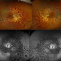 Retinitis Pigmentosa
Nov 7 2023 by Jolee Rodriguez
Bilateral fundus photography and fundus autofluorescence imaging of a 62-year-old male with Retinitis Pigmentosa. Patient reported visual field defects and dark adapting issues. Patient's vision at the time images were taken were sc20/20 of the right eye and sc20/25 of the left eye. Dr. Sutherland determined that based on the patient's lack of family history, the most likely route of inheritance is autosomal recessive.
Photographer: Jolee Rodriguez
Imaging device: Optos California RGB
Condition/keywords: autofluorescence imaging, fundus photography, hereditary retinal dystrophy, Optos, OPTOS CALIFORNIA RGB, retinitis pigmentosa, ultra-wide field imaging, Ultra-wide field retinal imaging, ultra-widefield image
-
 Macular Dystrophy
Oct 25 2023 by Zach Seim
Optos Fundus Autofluorescence of an 84 year old male with Macular Dystrophy. Patient presented with VA of sc CF at 3 feet. Genetic testing was performed to ensure that cause was not genetic.
Photographer: Zach Seim
Imaging device: Optos California
Condition/keywords: Autoflourescence, dystrophy, macular dystrophy, Optos, OPTOS CALIFORNIA, right eye, ultra-wide field imaging
 Loading…