Search results (51 results)
-
 Astrocytic Hamartoma
Feb 27 2025 by Daniel Davis, OCT-C
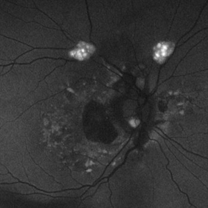
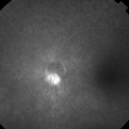
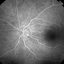
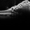
Fundus autofluorescence photo of 55-year-old female with astrocytic hamartoma in association with tuberous sclerosis. No treatment options available, benign. Other findings include; Posterior Vitreous Detachment, Vitreous Hemorrhage, Hereditary Retinal Dystrophy, Vitreous Opacities, Hypertensive Retinopathy.
Photographer: Daniel Davis, OCT-C
Imaging device: Optos California
Condition/keywords: astrocytic hamartoma, fundus autofluorescence (FAF)
-
 Astrocytic Hamartoma
Feb 27 2025 by Daniel Davis, OCT-C
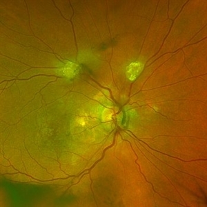
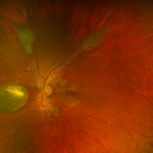
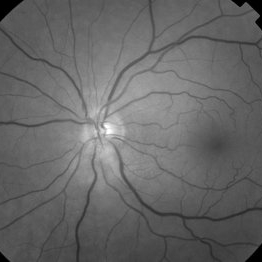
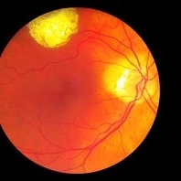
Color fundus photo of 55-year-old female with Astrocytic Hamartoma in association with tuberous sclerosis. No treatment options available, benign. Other findings include; Posterior Vitreous Detachment, Vitreous Hemorrhage, Hereditary Retinal Dystrophy, Vitreous Opacities, Hypertensive Retinopathy.
Photographer: Daniel Davis, OCT-C
Imaging device: Optos California
Condition/keywords: color fundus photograph
-
 Retinal Astrocytoma
May 9 2023 by JEFFERSON R SOUSA, Tecg.º (Biomedical Systems Technology)
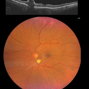
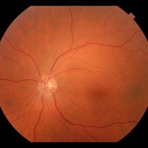
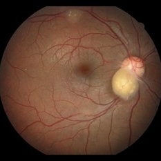
Retinal astrocytoma, also known as astrocytic hamartoma, is a rare benign tumor that occurs in the retina of the eye. It is a type of hamartoma, which means that it is made up of normal tissue that is growing in an abnormal way. Retinal astrocytomas are typically found in children and young adults and may be associated with a genetic condition called tuberous sclerosis complex. They can cause vision problems such as decreased visual acuity, visual field defects, and retinal detachment.
Photographer: JEFFERSON ROCHA DE SOUSA - Retinal Department at Institute Dr. Suel Abujamra Sao Paulo-Brazil
Imaging device: Clarus 700 - Zeiss, 135 degree images end CIRRUS 5000, Protocol, HD 5 Line
Condition/keywords: astrocytic hamartoma, astrocytoma
-
 Retinal Astrocytic Hamartomas
Dec 3 2019 by Timothy S Fuller, MD
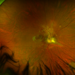
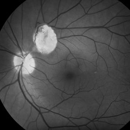
Fundus photograph of an 11-year-old boy with known tuberous sclerosis complex who was sent for evaluation of decreased vision in the right eye.
Photographer: Tony Abate
Imaging device: Optos
Condition/keywords: astrocytic hamartoma, tuberous sclerosis
-
 Retinal Astrocytic Hamartomas
Dec 3 2019 by Timothy S Fuller, MD
Fundus photograph of an 11-year-old boy with known tuberous sclerosis complex. Note calcification present in large lesion nasal to optic nerve.
Photographer: Tony Abate
Imaging device: Optos
Condition/keywords: astrocytic hamartoma, tuberous sclerosis
-
 Juxtapapillay Astrocytic Hemartoma with Moth Eaten Appearance on OCT
Oct 27 2019 by John S. King, MD
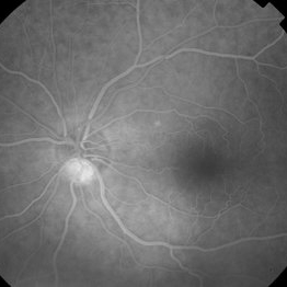
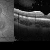
66-year-old white male without history of tuberous sclerosis was found to have an incidental, asymptomatic, translucent, retinal lesion with a few small telangiectatic vessels within it. The FA showed early hyperFL of these small vessels with prominent late leakage/staining. The OCT showed a retinal mass with a "moth eaten" appearance. Vision was 20/20 and the rest of the exam was unremarkable.
Photographer: Maisee Yang
Condition/keywords: astrocytic hamartoma
-
 2:30 FA - Astrocytic Hemartoma
Oct 27 2019 by John S. King, MD
66-year-old white male without history of tuberous sclerosis was found to have an incidental, asymptomatic, translucent, retinal lesion with a few small telangiectatic vessels within it. The FA showed early hyperFL of these small vessels with prominent late leakage/staining. The OCT showed a retinal mass with a "moth eaten" appearance. Vision was 20/20 and the rest of the exam was unremarkable.
Photographer: Maisee Yang
Condition/keywords: astrocytic hamartoma
-
 49 Sec FA - Astrocytic Hemartoma
Oct 27 2019 by John S. King, MD
66-year-old white male without history of tuberous sclerosis was found to have an incidental, asymptomatic, traslucent, retinal lesion with a few small telangiectatic vessels within it. The FA showed early hyperFL of these small vessels with prominent late leakage/staining. The OCT showed a retinal mass with a "moth eaten" appearance. Vision was 20/20 and the rest of the exam was unremarkable.
Photographer: Maisee Yang
Condition/keywords: astrocytic hamartoma
-
 28 Sec (Laminar Flow) FA - Astrocytic Hemartoma
Oct 27 2019 by John S. King, MD
66-year-old white male without history of tuberous sclerosis was found to have an incidental, asymptomatic, translucent, retinal lesion with a few small telangiectatic vessels within it. The FA showed early hyperFL of these small vessels with prominent late leakage/staining. The OCT showed a retinal mass with a "moth eaten" appearance. Vision was 20/20 and the rest of the exam was unremarkable.
Photographer: Maisee Yang
Condition/keywords: astrocytic hamartoma
-
 Red Free - Astrocytic Hamartoma
Oct 27 2019 by John S. King, MD
66-year-old white male without history of tuberous sclerosis was found to have an incidental, asymptomatic, translucent, retinal lesion with a few small telangiectatic vessels within it. The FA showed early hyperFL of these small vessels with prominent late leakage/staining. The OCT showed a retinal mass with a "moth eaten" appearance. Vision was 20/20 and the rest of the exam was unremarkable.
Photographer: Maisee Yang
Condition/keywords: astrocytic hamartoma
-
 FP - Astrocytic Hamartoma
Oct 27 2019 by John S. King, MD
66-year-old white male without history of tuberous sclerosis was found to have an incidental, asymptomatic, translucent, retinal lesion with a few small telangiectatic vessels within it. The FA showed early hyperFL of these small vessels with prominent late leakage/staining. The OCT showed a retinal mass with a "moth eaten" appearance. Vision was 20/20 and the rest of the exam was unremarkable.
Photographer: Maisee Yang
Condition/keywords: astrocytic hamartoma
-
 Retinal Phakomatous Hamartoma in Tuberous Sclerosis- Fundus Photos
Aug 28 2019 by Nisarg Joshi, MD
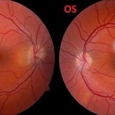
26-year-old female with history of tuberous sclerosis was found to have retinal phakomatous hamartomas in the right eye (peripapillary pale yellow lesion) and the left eye (opaque lesion in superior macula). The macula also shows RPE changes in both eyes.
Photographer: Nisarg Joshi, Geisinger Eye Institute, Danville, PA
Condition/keywords: hamartoma, phakoma, tuberous sclerosis
-
 Tuberous Sclerosis OCT Final
Aug 28 2019 by Nisarg Joshi, MD
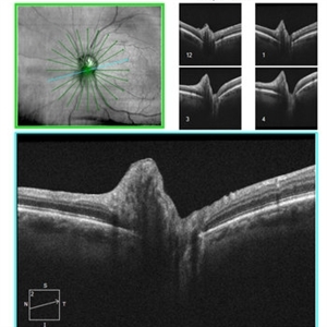
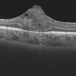
26-year-old female with history of tuberous sclerosis was found to have retinal phakomatous hamartomas in the right eye (peripapillary pale yellow lesion) and the left eye (opaque lesion in superior macula). This OCT shows the typical homogeneous appearance of these benign retinal lesions. The macula also shows RPE changes in both eyes.
Photographer: Nisarg Joshi, Geisinger Eye Institute, Danville, PA
Imaging device: Heidelberg Spectralis
Condition/keywords: hamartoma, phakoma, tuberous sclerosis
-
 Slide 4-42
Feb 20 2019 by Lancaster Course in Ophthalmology
Tuberous sclerosis. A later stage of the glial hamartoma. Small granules of calcium have developed as a degenerative change.
Condition/keywords: hamartoma, tuberous sclerosis
-
 Retina Hamartomas in Tuberous Sclerosis
Jan 8 2019 by Sofia Mano
Female 19-years-old with tuberous sclerosis. BCVA LE 10/10. Fundus LE shows three multinodular hamartomas.
Photographer: Sofia Sousa Mano
Imaging device: Canon CR 2 plus
Condition/keywords: hamartoma, tuberculous chorioretinitis
-
 Multiple Astrocytic Hamartomas
Jul 26 2018 by Olivia Rainey
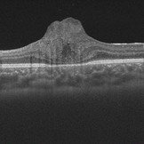
Optical coherence tomography of a 7-year-old female with multiple astrocytic harmartomas as a retinal manifestation of tuberous sclerosis. Patient came to our office to rule out possible drug toxicity from Sabril, a an anticonvulsant. There were no signs of retinal toxicity by extended ophthalmoscopy or imaging, yet she will be monitored every 6 months.
Photographer: Olivia Rainey
Imaging device: Heidelberg Spectralis
Condition/keywords: astrocytic hamartoma, Heidelburg Spectralis, infrared image, left eye, optical coherence tomography (OCT), tuberous sclerosis
-
 Astrocytoma OCT
Jan 9 2018 by Sidra Zafar
Swept source OCT imaging of retinal astrocytoma in a female child with known diagnosis of tuberous sclerosis.
Imaging device: Swept Source
Condition/keywords: astrocytoma, optical coherence tomography (OCT), tuberous sclerosis
-
 Astrocytoma OCT
Jan 9 2018 by Sidra Zafar
OCT imaging of retinal astrocytoma in a female child with known diagnosis of tuberous sclerosis. A cystic pocket can be observed.
Imaging device: Swept Source
Condition/keywords: tuberous sclerosis
-
 Retinal Astrocytic Hamartoma
Feb 11 2017 by Alexandra L Pappas, MD, MS
55-year-old male with an incidental finding. No other systemic findings of tuberous sclerosis.
Photographer: Cheri Scully, LPN
Condition/keywords: astrocytic hamartoma
-
 Astrocytic Hamartoma from Tuberous Sclerosis
Sep 18 2016 by John T. Thompson, MD
OCT of astrocytic hamartoma in child with tuberous sclerosis.
Imaging device: Heidelberg Spectralis
Condition/keywords: astrocytic hamartoma, tuberous sclerosis
-
 Hamartoma Tuberous Sclerosis
Jun 7 2016 by Nelson Chamma Capelanes, MD
Fundus photograph of an 52-year-old man with tuberous sclerosis and retinal hamartoma.
Photographer: Nelson Chamma Capelanes, Fundação Hilton Rocha, Promédica Indaiatuba, Brazil
Imaging device: Heidelberg Spectralis
Condition/keywords: hamartoma, tuberous sclerosis
-
 Hamartoma Tuberous Sclerosis
Jun 2 2016 by Nelson Chamma Capelanes, MD
Fundus photograph of an 52-year-old man with tuberous sclerosis and retinal hamartoma.
Photographer: Nelson Chamma Capelanes, Fundação Hilton Rocha, Promédica Indaiatuba, Brazil
Condition/keywords: tuberous sclerosis
-
 Astrocytic Hamartoma
Apr 30 2015 by Mariam A Al-Feky, MD
A 15-year-old boy with history of seizures controlled on treatment. C/O: OD painless DV 10/7 ago (accidental discovery) O/E: BCVA OD: 6/60 ,, OS 6/6. AS: NAD OU. Pupil: RRR no RAPD OU. Fundus examination OD showed a retinitis like lesion with an overlying corkscrew vessel well evident on FFA with late leakage and CSR and OCT through the retinitis like lesion shows diffuse hypereflective thickeninig in the superficial NFL. Thorough history taking revealed that patient has seizures and MRI lesions suggestive of tuberous sclerosis. So this is exudative hamartoma secondary to tuberous sclerosis with marked resolution after single IVI of Lucentis. Retinitis like lesion with corkscrew vessels in FFA is typical together with the homogenous hypereflective thickening in the NFL.
Photographer: Mariam AL-Feky
Imaging device: Optical coherence tomography
Condition/keywords: astrocytic hamartoma
-
 Astrocytoma
Feb 9 2015 by Patricia Araújo
Fundus photography of an 15-year-old boy with tuberous sclerosis.
Photographer: Dr Patricia Correa
Condition/keywords: astrocytic hamartoma, tuberous sclerosis
-
 Tuberous Sclerosis
Jan 8 2015 by H. Michael Lambert, MD
Color image of peripapillary astrocytic hamartoma.
Condition/keywords: tuberous sclerosis
 Loading…