 Initializing download.
-
 By Nisarg Joshi, MD
Florida Retina Institute
Co-author(s): Jonathan Tsui, Geisinger Eye Institute, Danville, PA. Steven Marks, Geisinger Eye Institute, Danville, PA - Uploaded on Aug 28, 2019.
- Last modified by Caroline Bozell on Aug 29, 2019.
- Rating
- Appears in
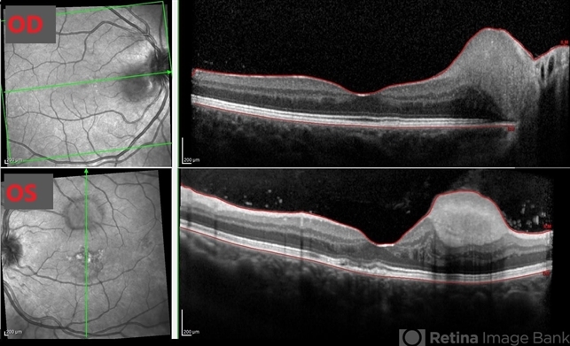
- Retinal Phakomatous Hamartomas in Tuberous Sclerosis
- Condition/keywords
- tuberous sclerosis, phakoma, hamartoma
- Photographer
- Nisarg Joshi, Geisinger Eye Institute, Danville, PA
- Imaging device
-
Optical coherence tomography system
Heidelberg Spectralis - Description
- 26-year-old female with history of tuberous sclerosis was found to have retinal phakomatous hamartomas in the right eye (peripapillary pale yellow lesion) and the left eye (opaque lesion in superior macula). This OCT shows the typical homogeneous appearance of these benign retinal lesions. The macula also shows RPE changes in both eyes.


