Search results (57 results)
-
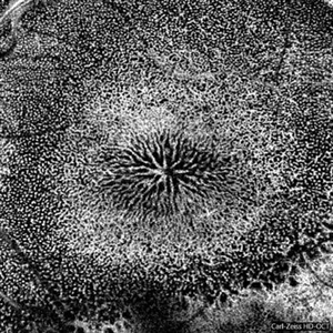
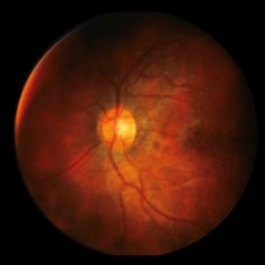
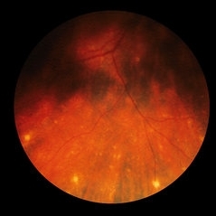
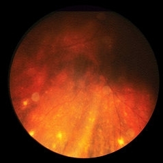
Ocular Toxocariasis
Jun 30 2025 by ASRS Staff
Ocular Toxocariasis found in a 46 year-old female patient with decreased vision. Findings were unilateral. Fundus photographs, echography and anterior segment ultrabiomicroscopy can be found at @eyemissu2
Condition/keywords: Ocular Toxocariasis
-
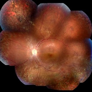
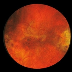
 Ocular Toxocariasis
Jul 4 2024 by Brandon I Fram, MD, BS
4 yo with toxocariasis-related peripheral granuloma with adhesion to the macula and macular subretinal fibrosis. Positive Toxocara titers.
Condition/keywords: toxocara canis, toxocara granuloma, toxocariasis
-
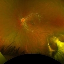
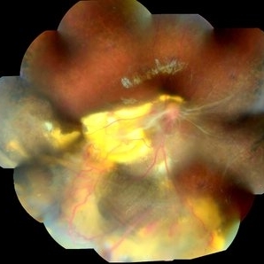
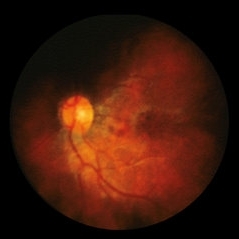
 Peripheral Toxocariasis Granuloma with Macular Traction
Jul 6 2021 by Lucas Zago Ribeiro, MD
15-year-old male patient with vision loss for the last 4 years, presenting peripheral toxocariasis granuloma, and macular traction.
Photographer: Lucas Zago Ribeiro, Federal University of São Paulo (UNIFESP)
Imaging device: Zeiss Visucam 524
Condition/keywords: granuloma, toxocariasis
-
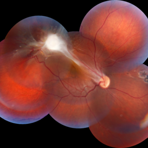
 Ocular Toxocariasis with Peripheral Granuloma
Apr 24 2021 by Alexandre Grandinetti, MD, PhD
8-year-old boy with a retinal fold secondary to peripheral toxocara canis granuloma localized on the superior retina.
Photographer: Corina Skrzek
Imaging device: Optos California
Condition/keywords: toxocariasis
-
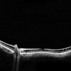
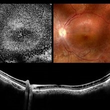 Retinoschisis
Mar 28 2021 by JEFFERSON R SOUSA, Tecg.º (Biomedical Systems Technology)
A 14-year-old male patient was admitted for visual evaluation. Visual acuity s/c in the right eye and 20/80 in the left eye. According to family members, he reported low vision since childhood. He had already undergone treatment with photocoagulation in another service to which he had a diagnostic hypothesis of Coats' disease. Laboratory tests were requested (HIV, TOXO, TOXOCARIASIS, ECA, VDRL, PPD). In the evaluation it was observed important exudation in the posterior pole, some vascular irregularities in the right eye. In the left eye, there is retinoschisis affecting the entire posterior pole and the region nasal to the optic disc, macula with a characteristic aspect of a cartwheel. Well exemplified by OCT-A (Structrure Deep: IPL - 25, OPL - 25).
Photographer: JEFFERSON R SOUSA - Study Center and Ophthalmological Research Dr. Andre M V Gomes, Institute Dr. Suel Abujamra São Paulo-Brazil
Imaging device: Optical Coherence Tomography system OCT CIRRUS 5000, Line Protocol, HD 21 line. Cirrus 5000 does not do a wide-angle tomographic image. This assembly was done manually with the junction of 11 lanes of 9mm each.
Condition/keywords: Coats' disease, retinoschisis
-
 Retinoschisis
Mar 28 2021 by JEFFERSON R SOUSA, Tecg.º (Biomedical Systems Technology)
A 14-year-old male patient was admitted for visual assessment. Visual acuity s/c in the right eye and 20/80 in the left eye. According to family members, he reported low vision since childhood. He had already undergone photocoagulation treatment at another service for which he had a diagnostic hypothesis of Coats' disease. Laboratory tests were requested (HIV, TOXO, TOXOCARIASIS, ACE, VDRL, PPD). In the evaluation, there was significant exudation in the posterior pole, some vascular irregularities in the right eye. In the left eye, there is retinoschisis affecting the entire posterior pole and the nasal region to the optic disc, macula with a characteristic chariot-wheel appearance, well exemplified by OCT-A (Structrure Deep: IPL - 25, OPL - 25).
Photographer: JEFFERSON R SOUSA - Study Center and Ophthalmological Research Dr. Andre M V Gomes, Institute Dr. Suel Abujamra São Paulo-Brazil
Imaging device: Optical coherence tomography system Optical Coherence Tomography system OCT CIRRUS 5000, Line Protocol, HD 21 line. Cirrus 5000 does not do a wide-angle tomographic image. (Structrure Deep: IPL - 25, OPL - 25).
Condition/keywords: Coats' disease, retinoschisis
-
 Retinoschisis
Mar 28 2021 by JEFFERSON R SOUSA, Tecg.º (Biomedical Systems Technology)
A 14-year-old male patient was admitted for visual evaluation. Visual acuity s/c in the right eye and 20/80 in the left eye. According to family members, he reported low vision since childhood. He had already undergone treatment with photocoagulation in another service to which he had a diagnostic hypothesis of Coats' disease. Laboratory tests were requested (HIV, TOXO, TOXOCARIASIS, ECA, VDRL, PPD). In the evaluation it was observed important exudation in the posterior pole, some vascular irregularities in the right eye. In the left eye, there is retinoschisis affecting the entire posterior pole and the region nasal to the optic disc, macula with a characteristic aspect of a cartwheel. Well exemplified by OCT-A (Structrure Deep: IPL - 25, OPL - 25).
Photographer: JEFFERSON R SOUSA - Study Center and Ophthalmological Research Dr. Andre M V Gomes, Institute Dr. Suel Abujamra São Paulo-Brazil
Imaging device: Topcon TRC-50 DX, Imaginet 4.0, angle de 50 graus. Flash 50w-s
Condition/keywords: Coats' disease, retinoschisis
-
 Coats' Disease
Mar 28 2021 by JEFFERSON R SOUSA, Tecg.º (Biomedical Systems Technology)
A 14-year-old male patient was admitted for visual evaluation. Visual acuity s/c in the right eye and 20/80 in the left eye. According to family members, he reported low vision since childhood. He had already undergone treatment with photocoagulation in another service to which he had a diagnostic hypothesis of Coatas disease. Laboratory tests were requested (HIV, TOXO, TOXOCARIASIS, ECA, VDRL, PPD). In the evaluation it was observed important exudation in the posterior pole, some vascular irregularities in the right eye. In the left eye, there is retinoschisis affecting the entire posterior pole and the region nasal to the optic disc, macula with a characteristic aspect of a cartwheel. Well exemplified by OCT-A (Structrure Deep: IPL - 25, OPL - 25).
Photographer: JEFFERSON R SOUSA - Study Center and Ophthalmological Research Dr. Andre M V Gomes, Institute Dr. Suel Abujamra São Paulo-Brazil
Imaging device: Topcon TRC-50 DX, Imaginet 4.0, angle de 50 graus. Flash 50w-s
Condition/keywords: Coats' disease, retinoschisis
-
 Retinoschisis
Mar 28 2021 by JEFFERSON R SOUSA, Tecg.º (Biomedical Systems Technology)
A 14-year-old male patient was admitted for visual assessment. Visual acuity without s / c in the right eye counts fingers and 20/80 in the left eye. According to family members, he reported low vision since childhood. He had previously been treated with photocoagulation at another service for which he had a diagnostic hypothesis of Coats' disease. Laboratory tests were requested (HIV, TOXO, TOXOCARIASIS, ECA, VDRL, PPD). In the evaluation, there was significant exudation in the posterior pole, some vascular irregularities in the right eye. In the left eye, there is a retinoschisis affecting the entire posterior pole and the nasal region to the optic disc, a macula with the characteristic aspect of a star. Well exemplified by OCT-A (Structure Deep: IPL - 25, OPL - 25).
Photographer: JEFFERSON R SOUSA - Study Center and Ophthalmological Research Dr. Andre M V Gomes, Institute Dr. Suel Abujamra São Paulo-Brazil
Imaging device: Optical Coherence Tomography system OCT CIRRUS 5000, Line Protocol, HD 21 line. Cirrus 5000 does not do a wide-angle tomographic image. This assembly was done manually with the junction of 11 lanes of 9mm each.
Condition/keywords: Coats' disease, retinoschisis
-
 Slide 3-18
Feb 20 2019 by Lancaster Course in Ophthalmology
Area of anterior segments of eye with Toxocara endophthalmitis, showing iris infiltration by inflammatory cells, ectropion uveae, and angle closed by anterior synechiae ( x12).
Condition/keywords: anterior synechiae, ectropion uveae, endophthalmitis, iris, toxocariasis
-
 Slide 3-16
Feb 20 2019 by Lancaster Course in Ophthalmology
Enucleated eye with Toxocara endophthalmitis, showing severe posterior inflammation with retinal detachment and disorganization.
Condition/keywords: endophthalmitis, enucleation, toxocariasis
-
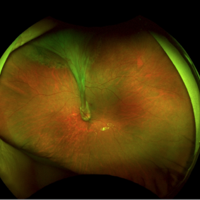
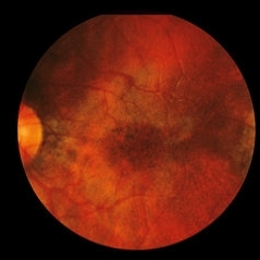
 Unilateral Subretinal Toxocara Granuloma
Apr 10 2018 by Albert Lin, MD
9-year-old male with chronic poor vision, band keratopathy, and posterior synechia in the right eye with a unilateral subretinal toxocara granuloma in the papillomacular bundle.
Photographer: Jody Watkins, University of Mississippi Medical Center, Department of Ophthalmology
Imaging device: Optos ultrawide field photo
Condition/keywords: toxocariasis
-
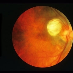
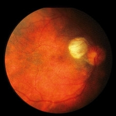
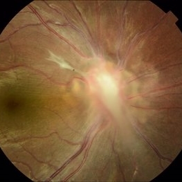 Ocular Toxocariasis
Feb 6 2018 by Bastián Schmidt Arias
These photographs show a classic localized granuloma located over the optic nerve.
Photographer: Bastian Schmidt
Imaging device: TRC-50DX - Topcon
Condition/keywords: toxocariasis
-
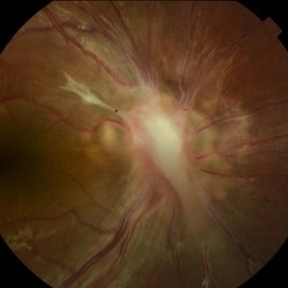 Ocular Toxocariasis
Feb 6 2018 by Bastián Schmidt Arias
These photographs show a classic localized granuloma located over the optic nerve.
Photographer: Bastian Schmidt
Imaging device: TRC-50DX - Topcon
Condition/keywords: toxocariasis
-
 Ocular Toxocariasis
Feb 6 2018 by Bastián Schmidt Arias
These photographs show a classic localized granuloma located over the optic nerve.
Photographer: Bastian Schmidt
Imaging device: TRC-50DX - Topcon
Condition/keywords: toxocariasis
-
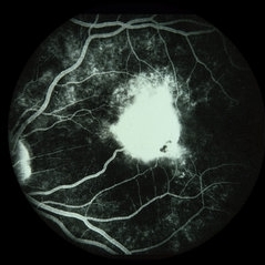 Toxocara Granuloma
Jun 4 2014 by Henry J. Kaplan, MD
Arteriovenous phase angiogram of the same patient shows staining of the granuloma and stippling hyperfluorescence around the lesion secondary to RPE window defect. #3
Condition/keywords: toxocara granuloma, toxocariasis
-
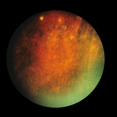 Cysticercus / Toxocara
Jan 15 2014 by David Callanan, MD
30-year-old female, HM; 20/20; later (2/1/90) 20/50 OD.
Condition/keywords: cysticercosis, toxocariasis
-
 Cysticercus / Toxocara
Jan 15 2014 by David Callanan, MD
30-year-old female, HM; 20/20; later (2/1/90) 20/50 OD.
Condition/keywords: cysticercosis, toxocariasis
-
 Cysticercus / Toxocara
Jan 15 2014 by David Callanan, MD
30-year-old female, HM; 20/20; later (2/1/90) 20/50 OD.
Condition/keywords: cysticercosis, toxocariasis
-
 Cysticercus / Toxocara
Jan 15 2014 by David Callanan, MD
30-year-old female, HM; 20/20; later (2/1/90) 20/50 OD.
Condition/keywords: cysticercosis, toxocariasis
-
 Cysticercus / Toxocara
Jan 15 2014 by David Callanan, MD
30-year-old female, HM; 20/20; later (2/1/90) 20/50 OD.
Condition/keywords: cysticercosis, toxocariasis
-
 Cysticercus / Toxocara
Jan 15 2014 by David Callanan, MD
30-year-old female, HM; 20/20; later (2/1/90) 20/50 OD.
Condition/keywords: cysticercosis, toxocariasis
-
 Cysticercus / Toxocara
Jan 15 2014 by David Callanan, MD
30-year-old female, HM; 20/20; later (2/1/90) 20/50 OD.
Condition/keywords: cysticercosis, toxocariasis
-
 Cysticercus / Toxocara
Jan 15 2014 by David Callanan, MD
30-year-old female, HM; 20/20; later (2/1/90) 20/50 OD.
Condition/keywords: cysticercosis, toxocariasis
-
 Cysticercus / Toxocara
Jan 15 2014 by David Callanan, MD
30-year-old female, HM; 20/20; later (2/1/90) 20/50 OD.
Condition/keywords: cysticercosis, toxocariasis
 Loading…