Search results (16 results)
-
 The Headlight in the Fog
Jun 17 2025 by Thirumalesh Mochi Basavaraj, MD
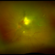
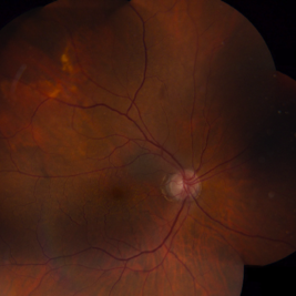
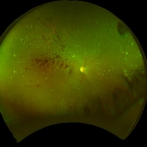
37 year old male with sudden onset diminution of visual acuity has a large retinochoridal granuloma along the superotemporal arcade and a few with satellite lesions more temporal to it, there was extensive Occlusoive vasculitis (both arterioles and veins )being involved with Vitrities.
Photographer: Vivekanand ,Narayana nethralaya
Imaging device: Daytona
Condition/keywords: acute toxoplasmosis, retinochoroiditis
-
 Toxoplasmosis
Dec 5 2024 by Tejaswita Verma
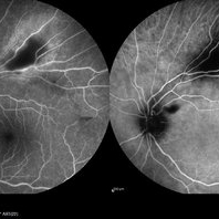
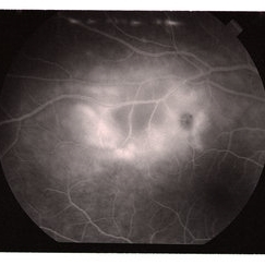
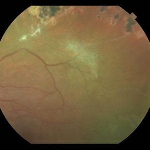
26 year old male with 6/18 vision , anterior chamber reaction, vitritis and retinitis lesion along the superotemporal arcade with full thickness involvement on OCT . FFA showing hypofluorescence with surrounding hyperfluorescence characterstic of toxoplasma retinitis . ICGA shows hypocyanescence.
Photographer: DR. TEJASWITA VERMA
Imaging device: MIRANTE
Condition/keywords: Fundus Fluorescein Angiography, indocyanine green (ICG) angiography, toxoplasmosis
-
 Toxoplasma Retinitis
Nov 4 2024 by Tejaswita Verma
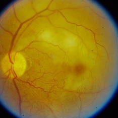
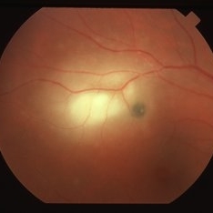
Fundus photograph of the left eye of a 20 year old male with 6/12 vision showing a posterior pole fluffy yellowish white lesion along superotemporal arcade with full thickness involvement of retinal layers on OCT suggestive of toxoplasma retinitis .He was started on Tab. Bactrim-DS followed by oral steroids after 4 days .There was anterior segment involvement with few Keratic precipitates , IOP was 47mm Hg, therefore patient was misdiagnosed as viral trabeculitis elsewhere. IOP was managed medically.
Photographer: DR. TEJASWITA VERMA
Imaging device: MIRANTE
Condition/keywords: toxoplasmosis retinitis
-
 Ruptured Retinal Arterial Macro-Aneurysm
Oct 27 2024 by César Adrián Gómez Valdivia, MD
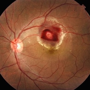
Ruptured retinal arterial macro-aneurysm found in a 56 YO female patient with history of untreated hypertension. Round or fusiform dilation of a retinal arteriole is usually seen within a third degree branch of one of the four main arcade arteries. Most common location for a symptomatic macroaneurysm is from a branch of the superotemporal arcade.
Photographer: @eyemissu2
Imaging device: TOPCON TRC-50DX
Condition/keywords: ruptured macroaneurysm
-
 Branch Retinal Artery Occlusion (BRAO)
Sep 26 2023 by Ben Serar
Fundus photograph of LE showing retinal edema and opacification along the superotemporal arcade, with cherry red spot at the macula, in a case of Branch Retinal Artery Occlusion (BRAO).
Condition/keywords: branch retinal artery occlusion (BRAO), cherry red spot
-
 Tributary vein occlusion
Sep 26 2023 by Ben Serar
Fundus photograph of LE showing retinal haemorrhages along the superotemporal arcade , with splinter haemorrhage near the disc, in a case of tributary vein occlusion.
Condition/keywords: Tributary vein occlusion
-
 CMV retintis
Sep 14 2023 by Ben Serar
Fundus photograph of LE showing superficial flame-shaped haemorrhages with surrounding retinal necrosis at the posterior pole along the superotemporal arcade in a case of fulminant type of CMV retinitis.
Condition/keywords: CMV retinitis, fulminant retinitis, pizza-pie appearance, viral retinitis
-
 Ruptured Retinal Macroaneurysm
Apr 26 2022 by Deepak Bhojwani, MS
Fundus image of a 58 year old hypertensive gentlemen with a ruptured macroaneurysm in superotemporal arcade along with massive intraretinal macular haemorrhage.
Photographer: DEEPAK BHOJWANI
Condition/keywords: arteriolar macroaneurysm, ruptured macroaneurysm
-
 Early Arterial to Venous Phase
Aug 26 2019 by Narciso F. Atienza, MD, MBA, FASRS, FPCS, FPAO.
Early arterial to venous phase shows beginning asymmetrical perfusion of the supero-temporal arcade supplying the macula. Infero-temporal arcade shows slight perfusion. More prominent choroidal flush noted
Photographer: Narciso F Atienza, Jr. MD, MBA
Imaging device: Topcon TRC
Condition/keywords: asymmetrical perfusion, superotemporal arcade
-
 02123-20190508-171643-Fluorescein-R-001
Aug 26 2019 by Narciso F. Atienza, MD, MBA, FASRS, FPCS, FPAO.
47-year-old female who came in with blurring of vision of the right eye of 2 weeks duration. She is hypertensive with poor control, taking Amlodipine irregularly. Denies any cardiac problem non-diabetic. Vision upon presentation was 20/400 (OD), 20/20 (OS) . Early arterial phase shows beginning asymmetrical perfusion of the supero-temporal arcade supplying the macula. Infero-temporal arcade shows no perfusion.
Photographer: Narciso F Atienza, Jr. MD, MBA
Imaging device: Topcon TRC
Condition/keywords: asymmetrical perfusion, inferotemporal arcade, superotemporal arcade
-
 Idiopathic Sclerochoroidal Calcification
Aug 12 2019 by Jonathan C. Tsui, MD
A 73-year-old Caucasian male presents with asymptomatic unilateral idiopathic sclerochoroidal calcification at the right superotemporal arcade. B-scan demonstrated hyperreflectivity of the lesion. Recent electrolyte testing was unremarkable.
Condition/keywords: idiopathic sclerochoroidal calcification
-
 Posterior Scleritis
Jun 6 2019 by Gary R. Cook, MD, FACS
Mid-phase fluorescein angiogram image of acute posterior scleritis lesion beneath the superotemporal arcade OD; V.A. = 20/40-2
Imaging device: Topcon VT-50
Condition/keywords: FA mid phase, fluorescein angiogram (FA), posterior scleritis
-
 Posterior Scleritis
Apr 2 2019 by Gary R. Cook, MD, FACS
White female with a focus of acute posterior scleritis beneath the superotemporal arcade OD; VA = 20/40-2
Imaging device: Topcon VT-50
Condition/keywords: posterior scleritis
-
 Lupus Hemorrhagic Occlusive Vasculitis
Apr 23 2018 by Frank Chin
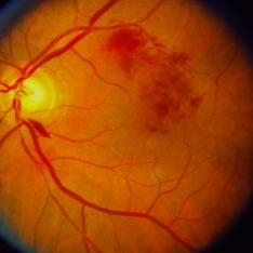
Fundus photograph of the right eye of a 24-year-old woman with history of systemic lupus erythematosus who presented with decreased visual acuity for 2-3 days found to have lupus hemorrhagic occlusive vasculitis with mild disc elevation, diffuse punctate cotton wool spots and dot blot hemorrhages, and a hemorrhage occlusive vasculitis along the superior branch of the superotemporal arcade involving the artery and vein.
Photographer: Frank Chin, MD, George Washington University
Imaging device: Optos 200Tx
Condition/keywords: blot hemorrhages, cotton wool spots, occlusive vasculitis, systemic lupus erythematosus (SLE) vasculitis
-
 Cytomegalovirus Retinitis Active
Sep 27 2012 by Jeffrey G. Gross, MD, FASRS
CMV retinitis active, superotemporal arcade.
Condition/keywords: superotemporal arcade
-
 Sickle Cell Retinopathy with Sea Fans
Aug 24 2012 by Geoffrey G. Emerson, MD, PhD, FASRS
Color fundus photograph of a 40-year-old man with African heritage and sickle SC disease. A sea fan (white) is present along the superotemporal arcade adjacent to an area of ischemia.
Photographer: Geoffrey Emerson, MD, PhD, Retina Center, Minneapolis
Condition/keywords: sea fan, sickle cell retinopathy
 Loading…