Search results (27 results)
-
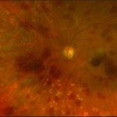
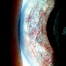
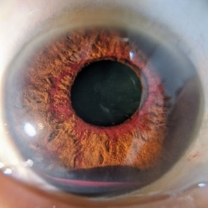 Rubeosis and Hyphema
Nov 19 2022 by Gareth Lema, MD, PhD
Florid rubeosis and hyphema.
Photographer: Gareth Lema, MD, PhD, New York Eye and Ear of Mount Sinai
Imaging device: Cell phone with a macro lens and muscle light for illumination.
Condition/keywords: Hyphema, Rubeosis
-
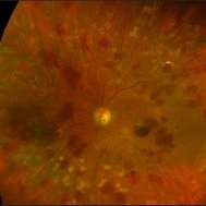
 Severe Rubeosis and Angle Neovascularization
Nov 21 2019 by Anfisa Ayalon, MD
Patient with proliferative diabetic retinopathy, severe retinal ischemia, rubeosis and angle neovascularization with IOP elevation.
Photographer: Anfisa Ayalon,MD., Meir Medical Center, Kfar Saba, Israel.
Imaging device: with gonioscopy lens
Condition/keywords: angle neovascularization, neovascular glaucoma, proliferative diabetic retinopathy (PDR), rubeosis
-
 Slide 12-25
Feb 27 2019 by Lancaster Course in Ophthalmology
Sequelae. The anterior chamber is deep, and the pupillary iris shows ectropion uveae resulting from shrinkage of new fibrovascular tissue (rubeosis iridis) on the anterior surface of the iris. The lens is dislocated posteriorly into the vitreous. All changes are the result of blunt trauma.
Condition/keywords: ectropion uveae, rubeosis, sequelae
-
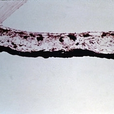 Slide 12-18
Feb 27 2019 by Lancaster Course in Ophthalmology
Rubeosis iridis. Fibrovascular tissue is present abnormally anterior to the anterior border layer of the iris. Shrinkage of such abnormal tissue results frequently in ectropion uveae (H&E xlOl).
Condition/keywords: fibrovascular tissue, rubeosis
-
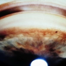 Slide 12-17
Feb 27 2019 by Lancaster Course in Ophthalmology
Rubeosis iridis. Vessels climbing over the anterior chamber angle have produced peripheral anterior synechiae.
Condition/keywords: rubeosis
-
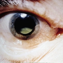
 Ocular ischemic syndrome
Jan 28 2018 by Alex H. Rubowitz, MD
A 65-year-old man with diabetes, presented with the typical round hemorrhages of OIS, as well as rubeosis iridis, and high intraocular pressure. Workup revealed carotid artery stenosis. Photos were taken just prior to his second laser PRP treatment.
Photographer: Lilach, Meir Hosopital Eye Clinic
Imaging device: Optos California
Condition/keywords: ocular ischemic syndrome
-
 OIS-Optos-photo-1
Jan 28 2018 by Alex H. Rubowitz, MD
A 65-year-old man with diabetes, presented with the typical round hemorrhages of OIS, as well as rubeosis iridis, and high intraocular pressure. Workup revealed carotid artery stenosis. Photos were taken just prior to his second laser PRP treatment.
Photographer: Lilach, Meir Hosopital Eye Clinic
Imaging device: Optos California
Condition/keywords: ocular ischemic syndrome
-
 Ocular Ischemic Syndrome
Jan 27 2018 by Alex H. Rubowitz, MD
A 65-year-old male with known diabetic retinopathy presented with iris rubeosis and neovascular glaucoma in his left eye. Photo was taken prior to his secind laser PRP session in the eye, and shows typical round hemorrhages of OIS. A systemic workup revealed severe carotid stenosis.
Photographer: Lilach, Meir Hospital Retina Clinic
Imaging device: Optos California
Condition/keywords: ocular ischemic syndrome
-
 Ocular Ischemic Symdrome
Jan 27 2018 by Alex H. Rubowitz, MD
A 65-year-old male with known diabetic retinopathy presented with iris rubeosis and neovascular glaucoma in his left eye. Photo was taken prior to his secind laser PRP session in the eye, and shows typical round hemorrhages of OIS. A systemic workup revealed severe carotid stenosis.
Photographer: Lilach, Meir Hospital Retina Clinic
Imaging device: Optos California
Condition/keywords: ocular ischemic syndrome
-
 Rubeosis Iridis
-
 Rubeosis Iridis
-
 Rubeosis following Ocular Ischemic Syndrome
Dec 10 2014 by Matt Poe, COA
Rubeosis following ocular ischemic syndrome.
Photographer: Matt Poe, COA. Northwest Arkansas Retina Associates, Springdale, AR.
Imaging device: Heidelberg HRA
Condition/keywords: ocular ischemic syndrome, rubeosis
-
---thumb.jpg/image-square;max$300,300.ImageHandler) DR Rubeosis
DR Rubeosis
-
---thumb.jpg/image-square;max$300,300.ImageHandler) DR Rubeosis
DR Rubeosis
-
---thumb.jpg/image-square;max$300,300.ImageHandler) DR Rubeosis
DR Rubeosis
Apr 4 2014 by H. Michael Lambert, MD
DR Rubeosis.
Photographer: Donald Lowd
Condition/keywords: diabetic rubeotic glaucoma, rubeosis
-
---thumb.jpg/image-square;max$300,300.ImageHandler) DR Rubeosis
DR Rubeosis
-
---thumb.jpg/image-square;max$300,300.ImageHandler) DR Rubeosis
DR Rubeosis
Apr 4 2014 by H. Michael Lambert, MD
DR Rubeosis.
Photographer: Donald Lowd
Condition/keywords: diabetic rubeotic glaucoma, rubeosis
-
---thumb.jpg/image-square;max$300,300.ImageHandler) DR Rubeosis
DR Rubeosis
Apr 4 2014 by H. Michael Lambert, MD
DR Rubeosis.
Photographer: Donald Lowd
Condition/keywords: diabetic rubeotic glaucoma, rubeosis
-
---thumb.jpg/image-square;max$300,300.ImageHandler) DR Rubeosis
DR Rubeosis
Apr 4 2014 by H. Michael Lambert, MD
DR Rubeosis.
Photographer: Donald Lowd
Condition/keywords: rubeosis
-
---thumb.jpg/image-square;max$300,300.ImageHandler) DR Rubeosis
DR Rubeosis
Apr 4 2014 by H. Michael Lambert, MD
DR Rubeosis.
Photographer: Donald Lowd
Condition/keywords: diabetic rubeotic glaucoma, rubeosis
-
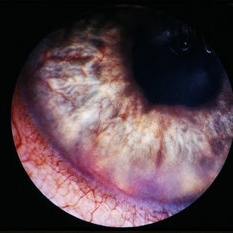
---thumb.JPG/image-square;max$300,300.ImageHandler) Rubeosis 1
Rubeosis 1
Jul 14 2013 by Jason S. Calhoun
Slit lamp photo shows active rubeosis in the left eye.
Photographer: Jason S. Calhoun, Department of Ophthalmology, Mayo Clinic Jacksonville, Florida
Imaging device: TOPCON D-90 SL NIKON CAMERA
Condition/keywords: rubeosis
-
 Rubeosis
Jul 14 2013 by Jason S. Calhoun
Slit lamp photo shows active rubeosis in the left eye.
Photographer: Jason S. Calhoun, Department of Ophthalmology, Mayo Clinic Jacksonville, Florida
Imaging device: TOPCON D-90 SL NIKON CAMERA
Condition/keywords: rubeosis
-
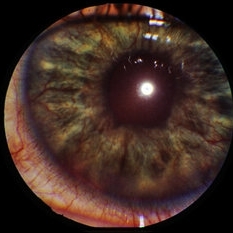
---thumb.JPG/image-square;max$300,300.ImageHandler) Rubeosis Iridis
Rubeosis Iridis
Jul 8 2013 by Jason S. Calhoun
Patient presents with rubeosis iridis in the right eye due to neovascular glaucoma. VA is 20/40 in the right eye. Will follow up in 3 months.
Photographer: Jason S. Calhoun, Department of Ophthalmology, Mayo Clinic Jacksonville, Florida
-
---thumb.JPG/image-square;max$300,300.ImageHandler) Rubeosis Iridis
Rubeosis Iridis
Jul 8 2013 by Jason S. Calhoun
Patient with rubeosis iridis in the right eye due to neovascular glaucoma. VA is 20/40 in the right eye. Will follow up in 3 months.
Photographer: Jason S. Calhoun, Department of Ophthalmology, Mayo Clinic Jacksonville, Florida
-
 Rubeosis Iridis
Jul 8 2013 by Jason S. Calhoun
Patient with rubeosis iridis in the right eye due to neovascular glaucoma. VA is 20/40 in the right eye. Will follow up in 3 months.
Photographer: Jason S. Calhoun, Department of Ophthalmology, Mayo Clinic Jacksonville, Florida
 Loading…