Search results (14 results)
-
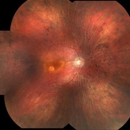
 Retinal Arteriolar Variation
Oct 31 2024 by AVIK DEY SARKAR, MS, FVRS, FAICO(VR)
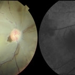
A 43-year-old hypertensive patient, diagnosed with Non-Ischemic Central retinal vein Occlusion in OS, presented with a striking anatomical variation in retinal vasculature. The inferior first-order retinal arteriole after initiating from the optic disc bifurcates, before reaching the fovea, and the superior branch after crossing the midline forms the superior arcade afterwards and produces dichotomous branching as usual. This defies basic anatomical considerations for retinal vasculature as they never cross the midline, also known as the watershed line for retinal vessels.1,2 References: 1. May CA, Rutkowski P. The Horizontal Raphe of the Human Retina and its Watershed Zones. Vision. 2019; 3(4):60. 2. May CA, Rutkowski P. Hypothesis: watershed zones in the human eye are a key for understanding glaucomatous retinal damage. Med Hypotheses. 2017;109:1-5.
Photographer: Dr. Avik Dey Sarkar, MBBS, MS, FVRS, FAICO, Consultant, Department of Vitreoretinal Services, Aravind Eye Hospital, Madurai, India
Imaging device: Wide angled Fundus imaging with Clarus 300
Condition/keywords: background diabetic retinopathy (BDR), Diabetic Retinopathy, retina, vascular anomaly
-
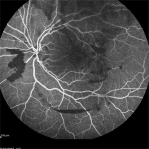
 From Ora to Ora
Aug 26 2024 by Nassim Alejandro Abreu Arbaje, MD
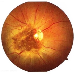
Ultra-wide field OCT angiography of a 39 year-old healthy male. The photo attempts to explore retinal vasculature up to the ora serrata.
Photographer: Johel Arrieta, TowardPi
Imaging device: TowardPi BMizar 400khz
Condition/keywords: OCT Angiography, OCTA, ultra-wide field imaging
-
 Ophthalmic Artery Occlusion in a 39-Year-Old with Rheumatoid Arthritis
Oct 6 2020 by Michael Izzo, MD
Left image: fundus photograph of a 39-year-old male with rheumatoid arthritis found to have ophthalmic artery occlusion depicting boxcar segmentation of blood in retinal vasculature and macular ischemia demonstrated by retinal whitening without cherry red fovea. Right image: early phase fluorescein angiography demonstrating patchy choroidal filling, arterial non-perfusion and optic nerve head leakage.
Photographer: Karen Rivera, COA; Washington National Eye Center
Condition/keywords: fluorescein angiogram (FA), ophthalmic artery occlusion, rheumatoid arthritis
-
 Posterior Vitreous Detachment
Sep 1 2020 by J. Sebag, MD, FACS, FRCOphth, FARVO
Left: Preset lens biomicroscopy of PVD in the left eye of a subject with a widely dilated pupil. The detached posterior vitreous cortex is seen (arrows) as is the optic disc and retinal vasculature (upper left). (courtesy of C. L. Trempe MD, Harvard Medical School, Boston, MA) [Sebag J: Vitreous – in Health & Disease Springer, New York, 2014; image © Springer Nature, reprinted with permission] Right: B-scan ultrasonography of PVD images the detached posterior vitreous cortex with a visible Weiss Ring.
Condition/keywords: posterior vitreous detachment
-
 Combined Hamartoma of the Retina and Retinal Pigment Epithelium
Apr 26 2020 by Dipak Nag, MBBS, FCPS, MSc, FRF
A 28-year-old female presented with a deeply pigmented gray- brown, elevated lesion extending from the temporal side of the disc to the macula (OU). Remarkable retinal vasculature with straightening of the distal vessels and dilatation as well as tortuousity of the peri-lesional vessels. The vitreoretinal interface shows gliosis and epi-retinal membrane (ERM) formation.
Photographer: Dipak Nag
Condition/keywords: hamartoma, retinal pigment epithelium
-
 Epipapillary Membrane
Apr 8 2019 by Gary R. Cook, MD, FACS
Epipapillary membrane and anomalous retinal vasculature OS in a middle-aged white male.
Condition/keywords: epiretinal membrane (ERM)
-
 Cancer-Associated Retinopathy (CAR)
Jun 30 2018 by Peter G. Hovland, MD, PhD
Mosaic fundus photograph of affected right eye of 56-year-old woman 3 years after onset of cancer-associated retinopathy. Demonstrates RPE atrophy and attenuated retinal vasculature. Patient presented with vitreous cells.
Photographer: Colorado Retina Associates
Condition/keywords: retinopathy, vitreous
-
 Cancer-Associated Retinopathy (CAR)
Jun 30 2018 by Peter G. Hovland, MD, PhD
Mosaic fundus photograph of affected right eye of 58-year-old woman 5 years after onset of cancer-associated retinopathy in fellow eye. Image demonstrates extensive RPE changes and attenuation of retinal vasculature.
Photographer: Colorado Retina Associates
Condition/keywords: retinal vasculature, retinopathy
-
 Sickle Cell Retinopathy
Sep 13 2015 by Thomas A. Ciulla, MD, MBA, FASRS
Angiography showed normal vessels posteriorly but severe capillary drop out throughout the periphery OU with scattered severe neovascularization at the edge of the capillary drop out peripherally. Note the preretinal and vitreous hemorrhage obscuring the view of the retinal vasculature.
Photographer: Thomas Steele
Condition/keywords: peripheral retinal neovascularization, sea fan, sickle cell retinopathy
-
 Fabry's Disease
Jun 4 2014 by Henry J. Kaplan, MD
Retinal vasculature tortuosity in Fabry's disease: OD. #2
Condition/keywords: Fabry disease
-
 Epiretinal membrane - Fundus photograph
Feb 5 2014 by Gerardo Garcia-Aguirre, MD
Fundus photograph of a 62 year old female with metamorphopsia and decreased visual acuity. A stage 2 epiretinal membrane is observed, causing distortion of the retinal vasculature.
Photographer: Gerardo Garcia-Aguirre, MD
Condition/keywords: epiretinal membrane (ERM)
-
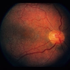
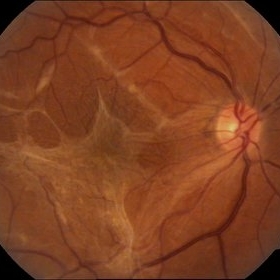
 Retinal Vascular Anomaly
Feb 20 2013 by From the Collections of Thomas M. Aaberg, MD and Thomas M. Aaberg Jr., MD
normal retinal vascular anomaly optic nerve vasculature
Condition/keywords: optic disc, retinal vasculature, vascular anomaly
-
 Oculocutaneous albinism Slide 2
Oct 22 2012 by Ronald C. Gentile, MD
Fundus photo of the left eye revealed absence of retinal and choroidal pigmentation with foveal hypoplasia. The choroidal vasculature was more prominent then the retinal vasculature.
Photographer: The New York Eye & Ear Infirmary Department of Medical Imaging
Condition/keywords: choroidal pigmentation with foveal hypoplasia, choroidal vasculature, retinal vasculature
-
 Regressed Proliferative Diabetic Retinopathy following PRP
Sep 6 2012 by Sharon Fekrat, MD FACS FASRS
58-year-old man with regressed proliferative diabetic retinopathy in the left eye following panretinal laser photocoagulation. Note attenuated retinal vasculature.
Photographer: Sarah Enfiedjian, Ophthalmic Photographer, Durham VA Medical Center, Durham, NC
Imaging device: Zeiss
Condition/keywords: attenuated vessels, pan-retinal photocoagulation (PRP)
 Loading…